Operator’s Manual 7 - 1
7
Strain Elastography
It is provided for reference, not for confirming a diagnosis.
It is produced based on the slight manual-pressure or human respiration in 2D real-time mode. The
tissue hardness of the mass can be determined by the image color and brightness. Besides, the
relative tissue hardness is displayed in quantitative manners.
7.1 Basic Procedure for Strain Elastography
Perform the following procedure:
1. Perform 2D scan to locate the region.
2. Tap [Elasto] to enter the elastography mode.
The system displays two dual B+E windows in real time. The left one is 2D image, and the
right one is elasto image.
3. Adjust ROI according to the lesion size.
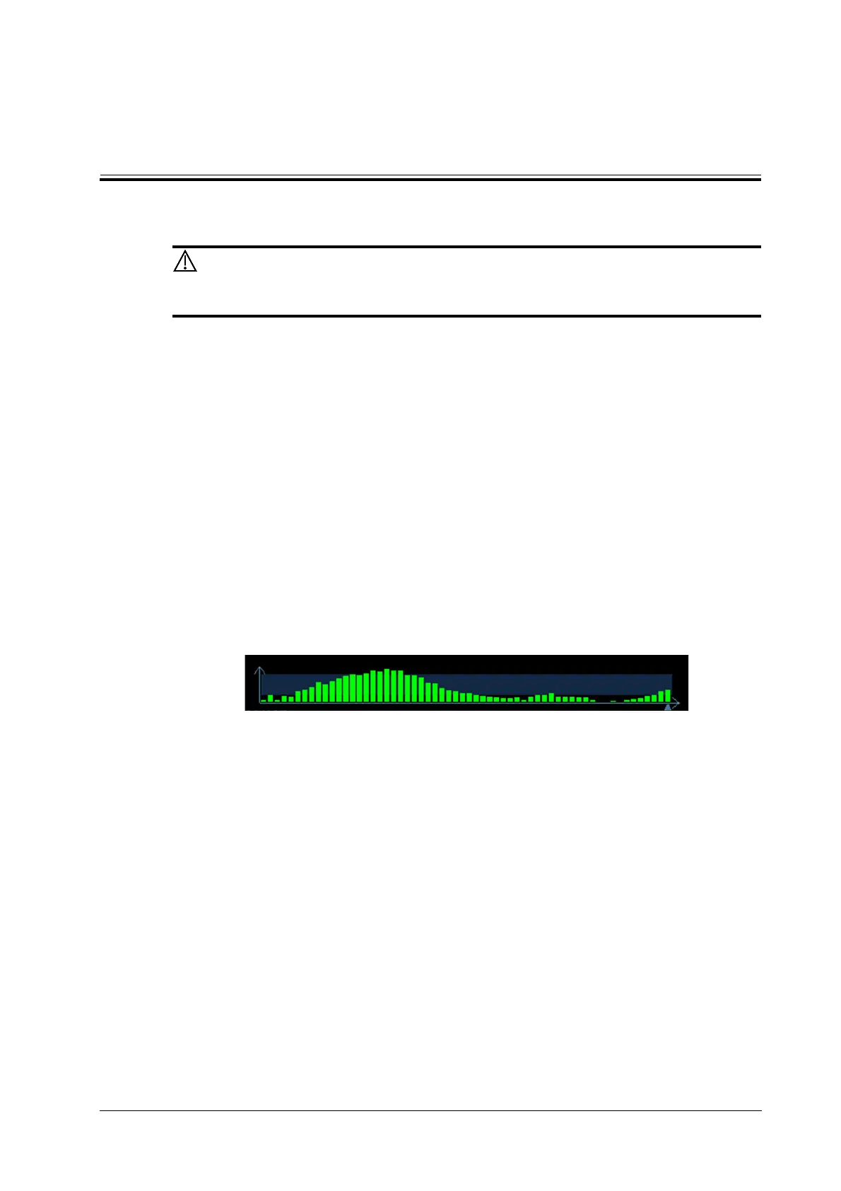
4. Press the probe according to the experiences and actual situation.
The screen displays the pressure curve in real-time:
Where, the X-axis represents time and Y-axis represents pressure.
5. Adjust the image parameters to obtain optimized image and necessary information.
6. Tap [B] or tap [Elasto] to exit, and then return to B mode.
7.2 Image Parameters
Smooth
Adjust the smooth feature of the Elasto image.
Opacity
Adjust the opacity feature of the Elasto image.
Invert
Invert the E color bar and therefore invert the colors of benign and malignant tissue.
Display Format
Adjust the display format of ultrasound image and the Elasto image.