74 300-002547-00 rev7
Axial, Sagittal, and Coronal CTs
The planar CT views are sized to fit the sidebar windows and zoom window. The
Monarch Navigation Application adaptively scrolls and pans the CT based on
scope location, keeping the scope always in frame.
The CT images are sized to fit the width of the window, scrolling only vertically.
When zoomed using the quick action button, the CT images scroll horizontally.
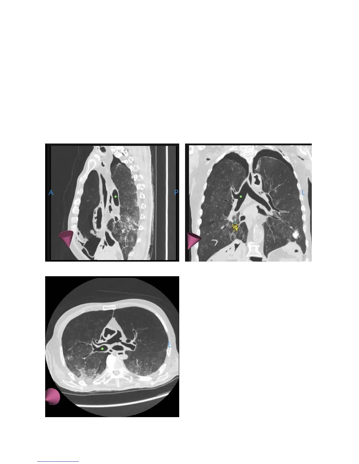
In the following images, the axial, sagittal, and coronal slices of the CT scan at
the location of the scope tip appear. In these images, the scope tip is
represented by a green ball and the direction of the tip is represented by a purple
cone.
Sagittal
Coronal