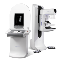
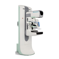
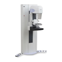
Selenia User Guide
Appendix A—System Specifications
122 MAN-03270 Revision 003
A.7 Imaging System Technical Information
A.7.1 Image Receptor
No fluid from incidental spillage on the top surface of the Image
Receptor seeps inside
Does not exceed 1.0 mm at maximum compression
24 cm x 29 cm Nominal. The active image area is marked on the
digital image receptor/breast platform cover.
Image Size, Screening and Diagnostic
Exams
18 x 24 cm nominal; locations: center, left, right 24 x 29 cm
nominal; center location only
Image Size, Diagnostic-Spot Compression
18 x 24 cm nominal; locations: center, left, right
Image Size, Diagnostic-Magnification
18 x 24 cm nominal; center location only
Digital Image Receptor MTF -Nyquist
frequency
50% or greater at 7.0 mR -0.0/+0.7 mR x-ray exposure
15% or greater at 7 mR -0.0/+0.7 mR x-ray exposure
X-ray exposure level at which image pixels are saturated is not
less than 1000 mR
Linear response over at least 400:1 in x-ray exposure
Lorad HTC™ high transmission cellular grid
The distance from the outside edge of the Image Receptor
enclosure to the Active Image Area along the chest wall is less
than 5 mm.
The distance from the outside edge of the detector enclosure to
the active detector area along the edges perpendicular to the
chest wall is less than 40 mm.
ACR Phantom Score at MGD = 2mGy
At least 5 fibers, 4 specks, 4 mass
The time between completion of an x-ray exposure and
availability of the Preview image: less than 20 seconds.
Within Federal Regulatory limit for screen-film mammography
systems (21CFR 1020): 0.1 mR/h.