Structure and Assembly/Disassembly 9-5
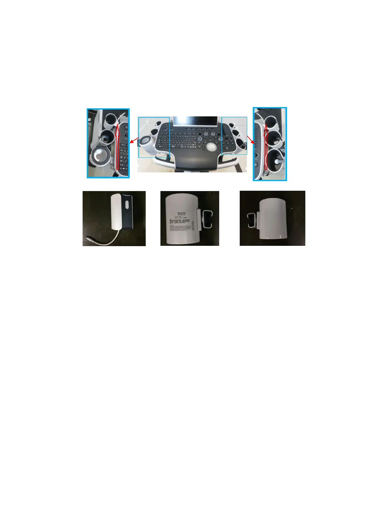
9.3.1 Large/Small Probe Holders, Left Bracket of Coupling
Gel Heating cup, Intracavitary Probe Holder
1. Remove the probe holder in the order of 4D probe, large probe holder, coupling gel heating cup
and small probe holder along the arrow’s direction.
2. Unscrew 2 M4 X 12 cross panhead screws with screwdriver (M3, M4) to remove intracavitary
probe bracket.
arge probe
eft bracket of
coupling gel
mall probe
arge probe
Coupling gel heating
cup 12V 12W
eft bracket of the
ht bracket of the
oupling gel heating cup

 Loading...