Structure and Assembly/Disassembly 9-29
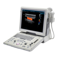
9. Buckle the display cable cover to the rear cover of the display along the direction shown in the
figure, use two M4x12 screws to fix it, and install the screw caps.
9.3.13 LCD Signal Connector PCBA assembly
1. Refer to 9.3.6 Control Panel Assembly for details.
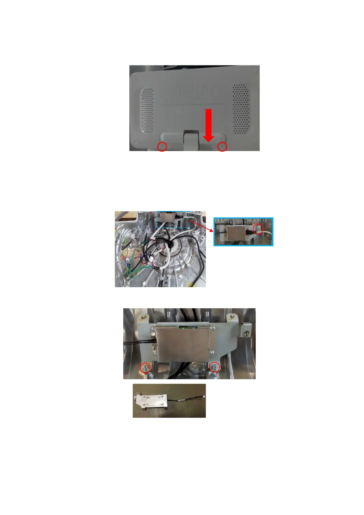
2. Unscrew M4 X 12 cross panhead screw on UC-0.5 lock knob with screwdriver (M3, M4) to
remove the signal cable of the monitor.
3. Unscrew 2 M4 X12 cross panhead screws with screwdriver (M3, M4) to remove the LCD signal
connector PCBA assembly.
signal connector PCBA assembly
 Loading...