Electrode Placement 4 ECG and Arrhythmia Monitoring
51
Electrode Placement
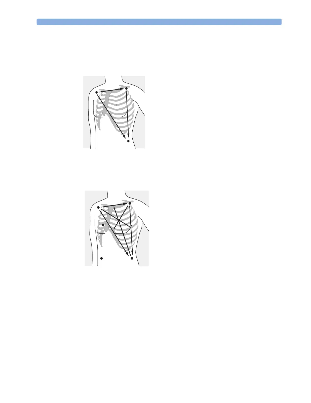
Figure 32 shows the typical electrode placement for a 3-lead ECG set.
Figure 32 3-lead Placement
Figure 33 shows the typical electrode placement for a 5-lead ECG set.
Figure 33 5-lead Placement
–
–
–
+
+
+
I
II
III
LA/L
(Black/
Yellow)
LL/F
(Red/
Green)
RA/R
(White/
Red)
RA/R placement: directly below the clavicle and
near the right shoulder
LA/L placement: directly below the clavicle and
near the left shoulder
LL/F placement: on the left lower abdomen
–
–
–
+
+
+
I
II
III
aVR
aVL
aVF
LA/L
(Black/
Yellow)
LL/F
(Red/
Green)
RL/N
(Green/
Black)
V/C
(Brown/
White)
RA/R
(White/
Red)
RA/R p
acement:
irect
y
e
ow t
e c
avic
e an
near
the right shoulder
LA/L placement: directly below the clavicle and near
the left shoulder
RL/N placement: on the right lower abdomen
LL/F placement: on the left lower abdomen
V/C placement: on the chest; the position depends on
your required lead selection. See Figure 34.
 Loading...