Chapter H - CEPHALOSTAT
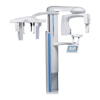
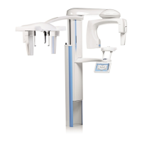
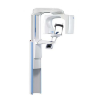
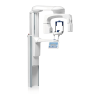
H-22 ProMax X-ray unit with DImax3
ADJUSTMENTS AND CALIBRATIONS
Technical manual
The
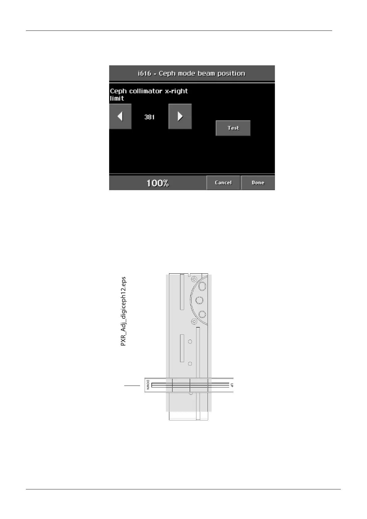
Ceph mode beam position
display appears. The sensor head and the first primary colli-
mator will move to the centre position. Touch the Test field.
Figure 37
Place the beam alignment tool to the sensor alignment tool to horizontal position
(see Fig. 38 below).
Stand behind the tube head and protect yourself from radiation. Press and hold down the
exposure button. The image of the radiation beam will appear on the alignment tool.
The radiation beam must appear symmetrically on the beam alignment tool as shown on
the Fig. 38 below. If it does not adjust the first primary collimator horizontal position.
Figure 38
Adjust the limit values with the arrow fields on the display and drive the primary collimator to
the selected position by touching the Test field. When the beam is correctly positioned, touch
the Done field.
Beam alignment tool
 Loading...