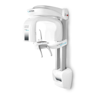
User Manual – General instructions for use
USER MANUAL • X-MIND prime • (19) • 01/2020 • NXMPEN010C
8.8.2.1
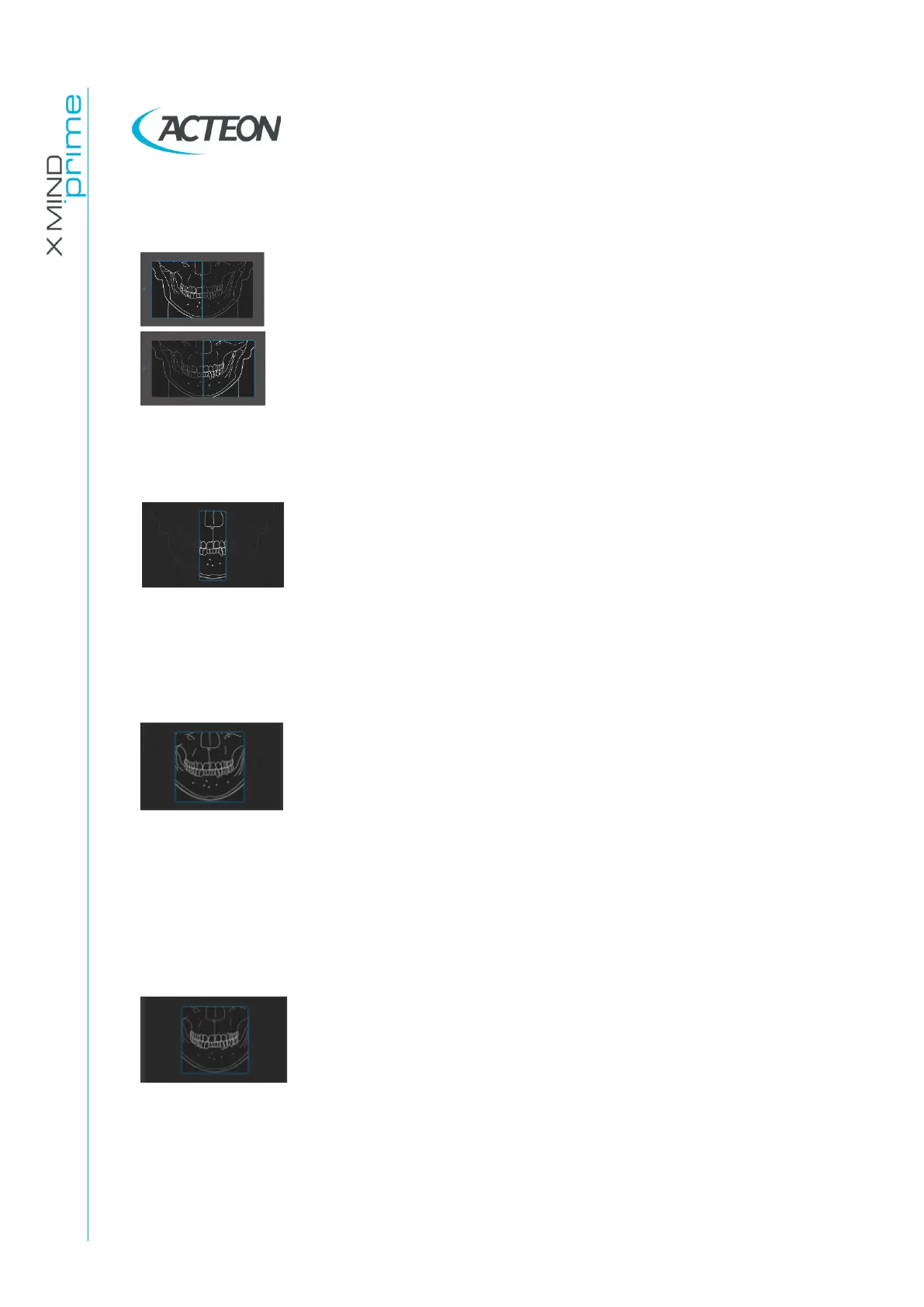
Left / Right Half Panoramic
The Half Panoramic mode, right or left, means that only the
corresponding half arch is irradiated; emission will start from the
beginning, to just after the mid sagittal plane for the right part. For
the left, it will start just before the mid sagittal plane and continue
until the end of the rotation.
These two kinds of exams are normally used when it is already
known that the patient has a problem on only one half of the arch,
so it is possible to reduce patient irradiation. Follow the
instructions for normal panoramic exams for patient positioning.
8.8.2.2
Frontal dentition
The Frontal dentition exam takes an X-ray of the frontal
dentition area (roughly from canine to canine). Follow the
instructions for normal panoramic exams for patient positioning.
8.8.2.3
Low dose Panoramic
The low dose panoramic exam takes an X-ray only of the dental
arch, excluding the ascending rami of the temporo-mandibular
joint from the image; the exam is performed with the same
trajectory of the standard Panoramic exam, reducing the rays'
emission time.
This exam is used, for instance, during treatment continuation
phases or where a lack of pathologies of the same joint is already
known.
Follow the instructions for normal panoramic exams for patient
positioning.
8.8.2.4
Ortho Rad dentition
The ortho rad panoramic exam delivers an image of the pure
dental arch, excluding the ascending rami branches of the
temporo-mandibular joint from the image; the trajectory of the
rotating arms is, however, optimised for a better orthogonality
between the X-ray beam and incident sections of near teeth.
Thus the image has reduced teeth overlapping, improving the
diagnosis of interproximal decay.
As a consequence of the different trajectory, the focus layer,
mainly in the front teeth area, is smaller and patient positioning
for this exam needs more care. Follow the instructions for normal
panoramic exams for patient positioning.