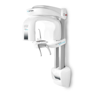
8.10 TMJ exam
The TMJ exam with open/closed mouth is similar to panoramic exams; the only
difference is that exposure is only on the TMJ (Temporo Mandibular Joint) area. The
operating sequence of the exam is therefore identical to that described for panoramic
exams.
The temporo-mandibular joint exam uses projection geometry giving an image of the X-
rayed condyle along a direction almost parallel with its major axis, in order to achieve a
clear view of its positioning inside the cavity.
The TMJ Standard function makes it possible to obtain 4 different acquisitions on the
same image, by performing two rotational movements. The 4 images represent the right
and left condyle of the temporo-mandibular arch (TMJ) with closed mouth and open
mouth.
Figure 25 shows the information related to the single sectors.