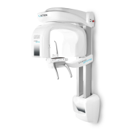
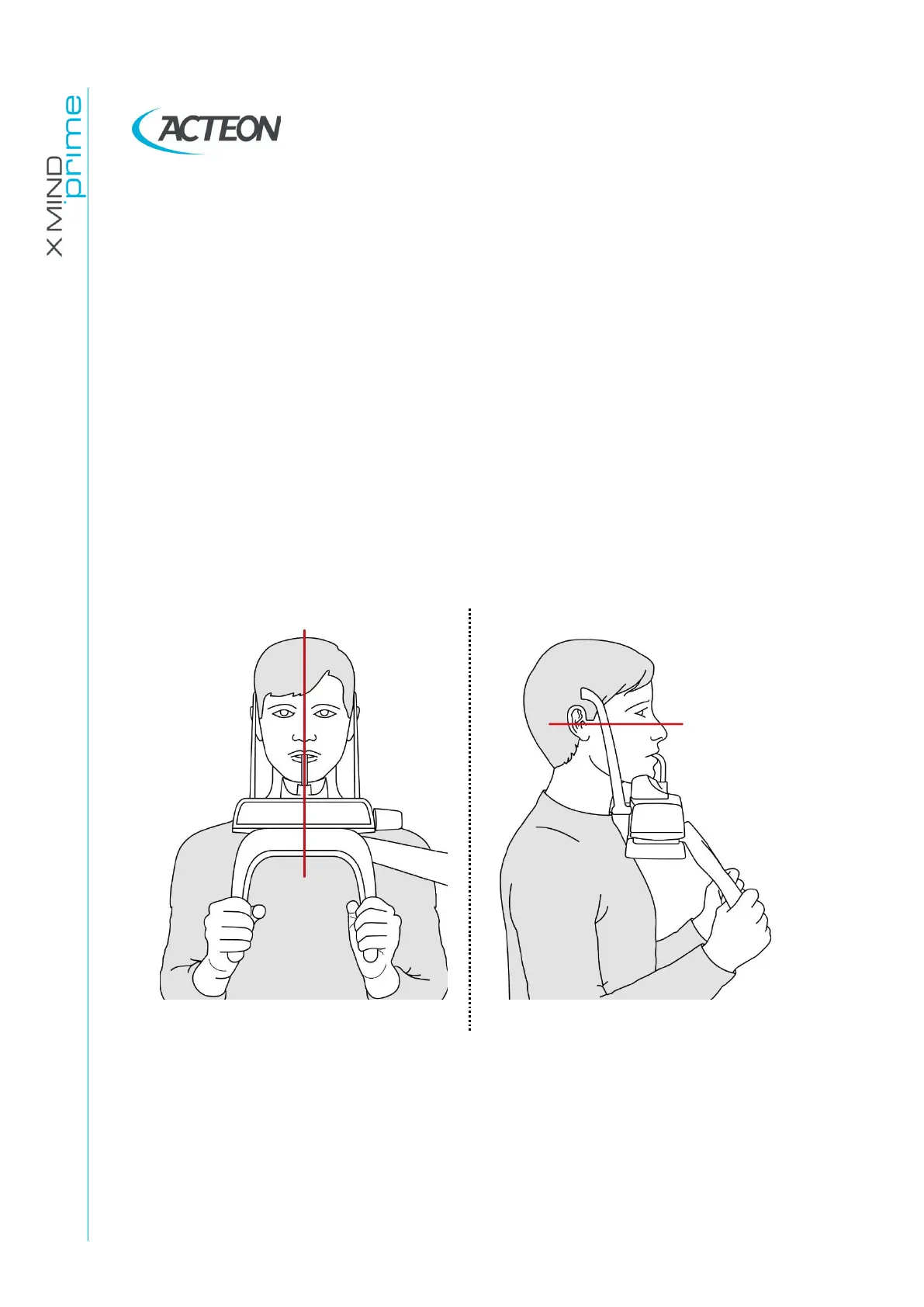
11.1 Proper positioning of the patient
Patient positioning is determining to get good quality radiography. This is due to the fact
that the shape of the focussed area, e.g. of the layer clearly shown on the image, tends
to follow the dental arch and has a non-constant deepness. The objects outside this
focused area will therefore appear blurred on the radiography.
1. The patient should not wear clothes that may interfere with the X-ray beams, also to
leave more space between the patient’s shoulders and the rotating arm of the
equipment. Care must be taken in order to avoid interference between the X-ray beam
and the protective apron worn by the patient.
2. Metal objects (necklaces, earrings etc.) must be avoided; these objects not only
create radio-opaque images in their own position, but also false images projected in
other parts of the radiography, so disturbing the correct view of the anatomy.
3. Patient’s incisors must be positioned into the reference notch of the bite.
4. Frankfurt plane (plane passing through the inferior margin of the orbit and the upper
margin of the ear canal) must be horizontal.
5. Mid-Sagittal plane must be centered and vertical.
Figure 32
6. Spine should be well stretched, this is normally obtained by asking the patient to
step forward, making sure that all other conditions are unchanged. If not properly
extended, the spine will cause the appearing of a lower exposed area (clearer) in
the front part of the image.