7. Instruct the patient to swallow and keep the tongue against the palate. Patient's
tongue must be held closely to the roof of the mouth during the exposure, otherwise
a dark air space between the dorsum of the tongue and the palate could obscure
the apical region of the maxillary teeth.
8. Patient must stay motionless during the examination.
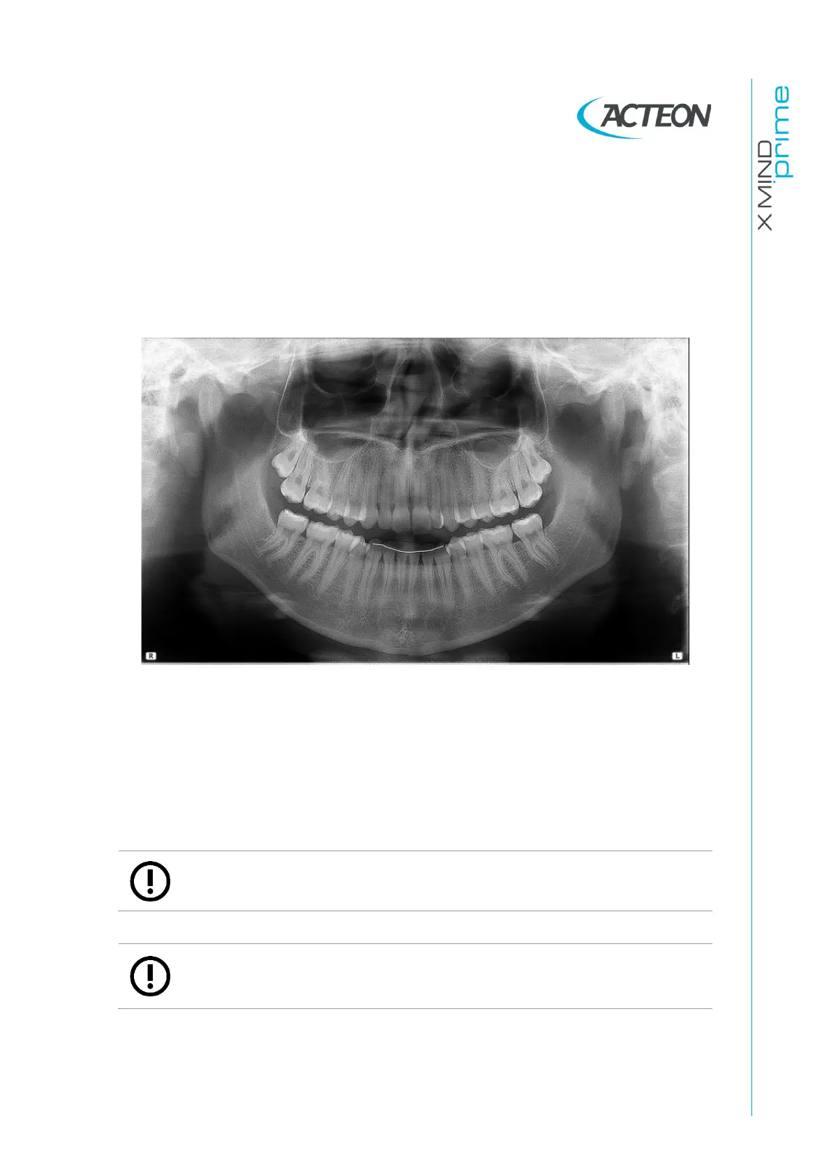
The result of all the above listed actions will be a radiography where all the parts are
properly exposed and are well identifiable as shown in Figure 33.
Figure 33
In a good panoramic image, all anatomic structures are well represented and an equal
magnification and sharpness of all structures can be seen.
The image must be symmetric, with the ascending rami of the temporo mandibular joints
almost parallel and showing posterior vertical borders. The occlusal plane is quite
smiling, despite this the palatal plane does not overlap the apex of the upper arch and
therefore allows a good view of the apex itself. The spine is well compensated.
Note
The region of the incisors is the most critical because the anterior portion of the
image layer is very narrow. Points 3 and 4 are determining for a good result.
Note
Any flaring of dentition may not allow crowns and apices of both arches to fit in
the image layer at the same time. For these patients, you must purposely move
him/her further forward in order to move the apices into the image layer.