Image Optimization 5-149
and measures the changes of IVC inner diameter. It can be used to assist in treatments, such as
volume estimation and fluid infusion.
NOTE: 1. The cardiac package should be configured in advance.
2. Smart IVC is an option.
3. Smart IVC supports calculation in both real time and freeze modes.
– Enable Smart IVC in real time. The calculation starts from the current frame
and ends after the image is frozen.
– Enable Smart IVC in freeze mode or from the cine file. The calculation starts
from the current frame and ends at the last frame. If the cine length is no
longer than 10 seconds, it is allowed to calculate in retrospective from the
current frame after the cine length reaches 10 seconds.
Perform the following procedure:
1. Select an appropriate probe and exam mode.
2. Move the probe to gain an appropriate IVC long axis image.
3. Tap [Smart IVC] on the touch screen or the user-defined key to enter Smart IVC mode and start
calculation.
a) Tap to select a breath type: Spontaneous Breath or Mechanical Ventilation.
b) Select [Change Resp time] > [[Resp Time(rpm)]] to set a respiratory time.
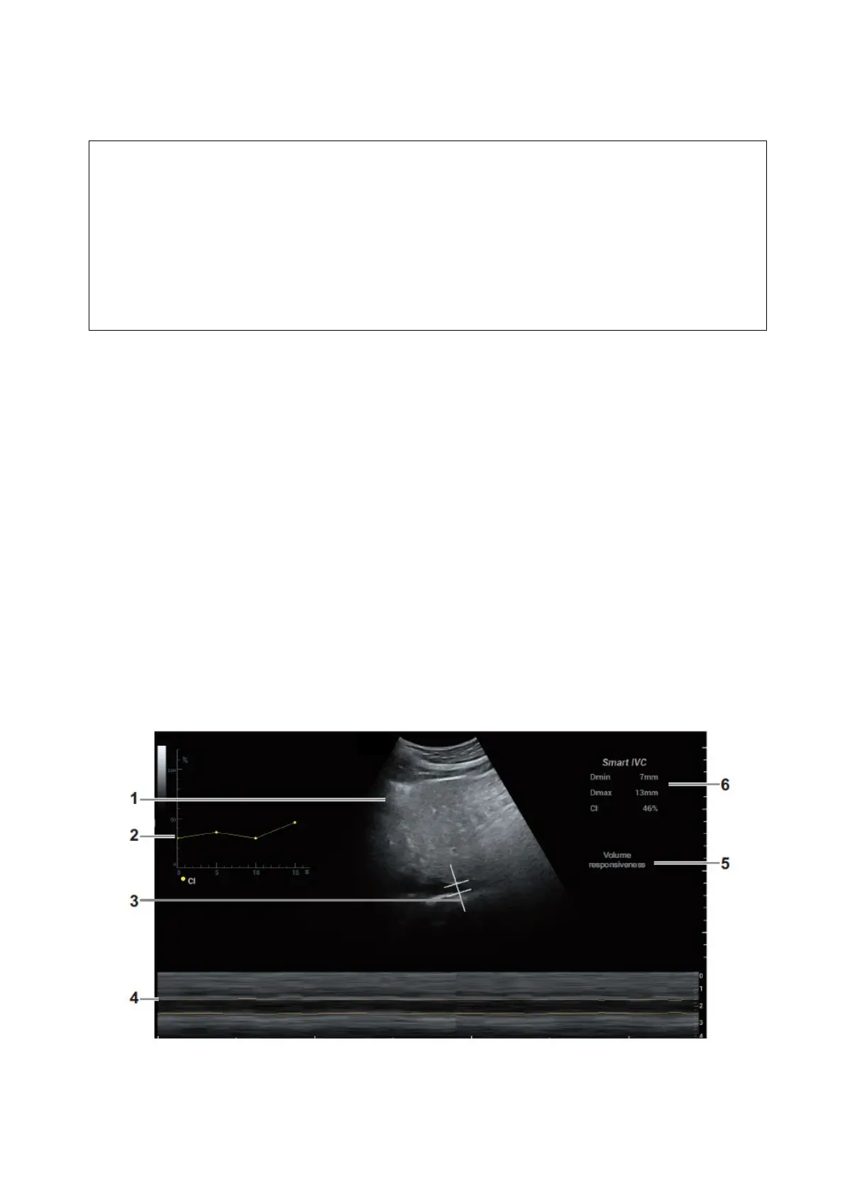
The system measures the IVC inner diameter of the image in every frame, calculates the maximum
and minimum IVC diameters, and draws a quantitative index change curve in real time.
4. If necessary, you can adjust the IVC sampling line manually:
a) Tap [Edit Line] on the touch screen.
b) Tap [Angle] on the touch screen to adjust the sampling line angle, and use the trackball to
adjust the sampling line position.
c) Press the <Update> key to start calculating IVC again.
5. Press the <Freeze> key to freeze the image and finish calculating IVC.
The calculation results and quantitative trend curve are displayed on the main screen. Tap
[Diagnostic Info] on the touch screen to add diagnostic information to the image quickly.
 Loading...


