5-20 Image Optimization
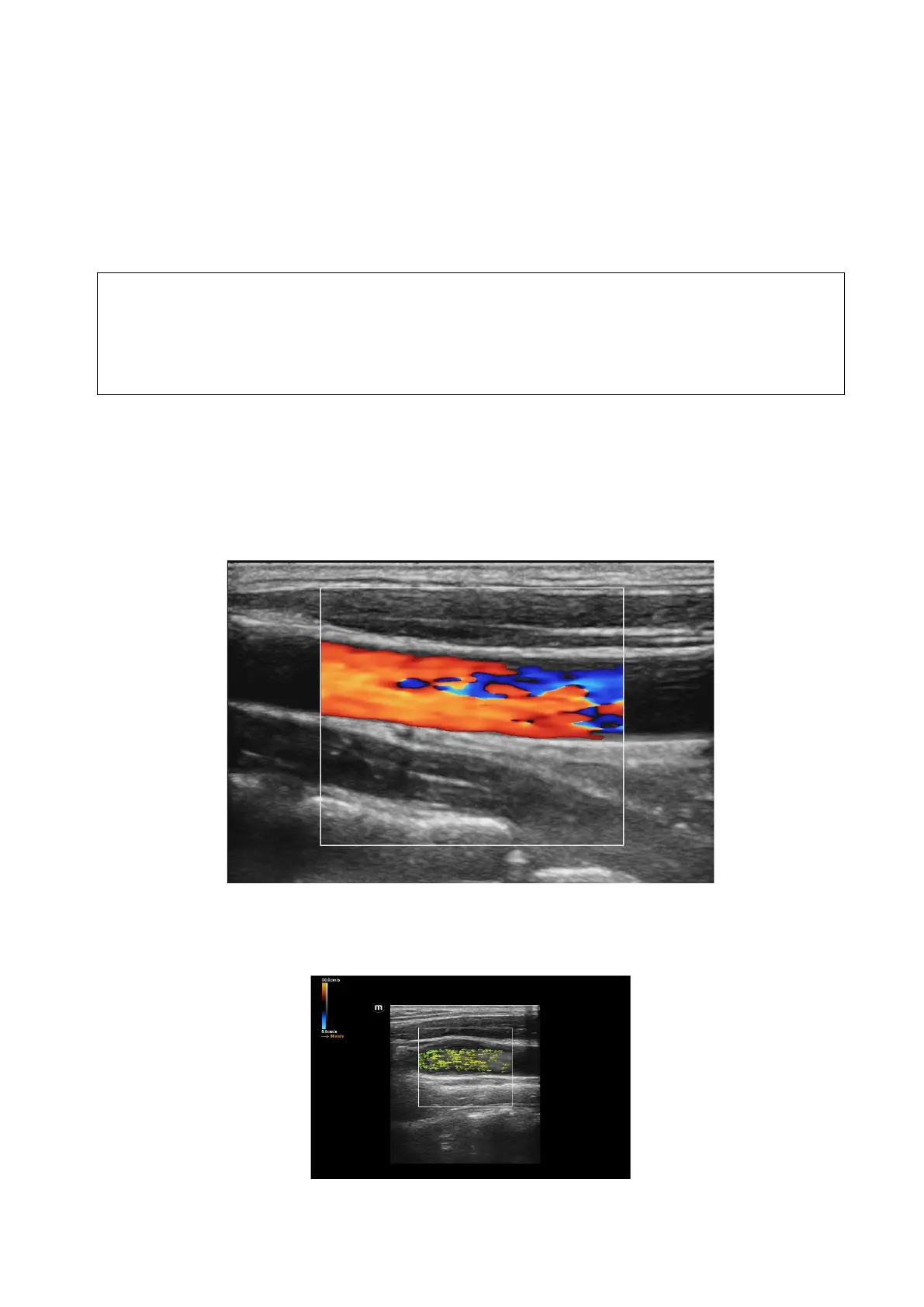
5.6 V Flow
V Flow shows the blood direction and velocity via the arrow. The arrow length represents the velocity,
and the arrow orientation represents the direction of the blood flow. The blood flow is displayed
according to the updates of the arrow position and the velocity.
V Flow shows the blood situation on vortex flow, turbulent flow, regurgitation, etc.
Description 1. V Flow imaging is an option.
2. You can enter V Flow on B/Color real-time scan mode.
3. It only supports single-window display, but does not support dual-window display
and quad-window display.
4. V Flow image does not support the image magnification.
5.6.1 V Flow Basic Operations
1. Scan the carotid artery on the real-time B/Color mode; adjust the probe and the image to locate the
desired region at the center of the B/Color mode image.
2. Tap [V Flow] or user-defined key to enter V Flow mode. Roll the trackball to locate the ROI, and
press <Set> and roll the trackball to adjust the ROI position and the size.
3. To optimize the image, adjust the parameters during the image scan. See Chapter 5.12.3 Cine
Review for details.
4. Tap [Start Capture] or press <Update>, and keep the probe still. The image shows the change of
the blood flow over a period of time.
 Loading...


