5-142 Image Optimization
5.17.12 Measuring
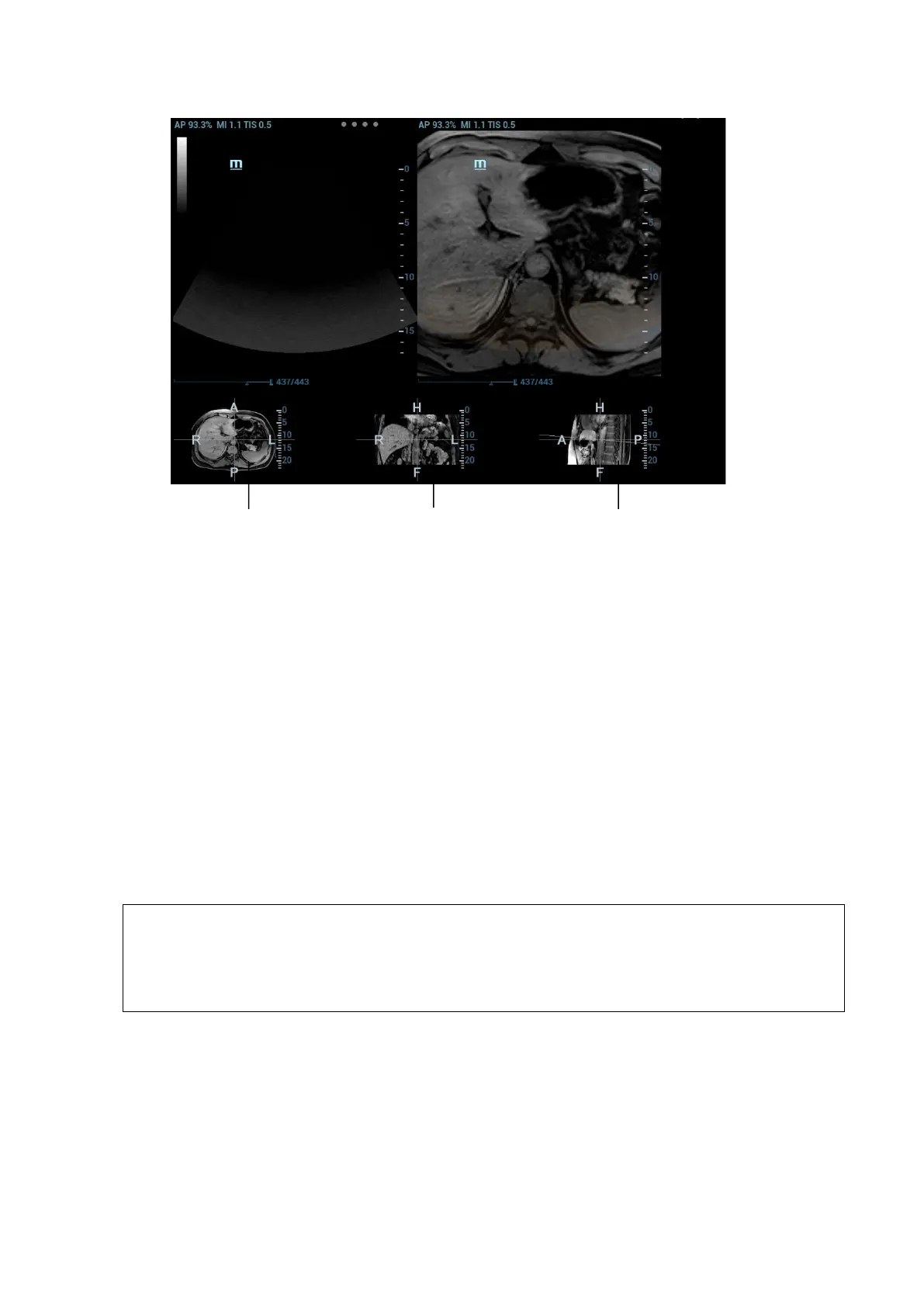
It is available to conduct the general measurements on the image that the ultrasound image registers
with the CT/MR/PET/freehand image. See also Chapter 8.2 General Measurements.
5.17.13 Comment and Body Mark
It is available to conduct the comment and the body mark on the image that the ultrasound image
registers with the CT/MR/PET/freehand image. See also Chapter 9.1 Comments.
5.18 RIMT (Real-time Intima-Media Thickness)
RIMT early detects and prevents coronary heart disease, artery vessel from being pathological, and
estimates the therapeutic effects. RIMT detects the changes of the vascular intima in real time, and monitors
and calculates the thickness of the carotid intima automatically.
Note
RIMT is an option.
It is merely available to enter RIMT imaging mode in B single window and dual
window when adopting linear probe for carotid exam.
Do not press the probe after entering RIMT imaging mode when scanning the image in
real time.
1. Select the probe first. Perform B mode in carotid exam mode. Detect the patient’s carotid in B
mode. Keep the acoustic beam vertical with the anterior and the posterior of the vascular and make
the anterior and the posterior of the intima visible at the carotid stenosis to obtain a premium image.
2. Tap [RIMT] to activate the function. Rotate [Side] to select left or right carotid.
3. Roll the trackball to locate ROI over the target area. The dotted line of the ROI is in the middle of
the blood vessel. Press <Set> to confirm the position and size of the ROI.
Transverse plane
Coronal plane
Sagittal plane
 Loading...


