USER'S MANUAL
General instructions for use
I-Max Touch (110-120V) (Rev. 0)
108
6.12.1 Proper positioning of the patient
The proper positioning of the patient during the panoramic examination
is very important in order to get a good quality radiography. This is due
to the fact that the shape of the focussed area, e.g. of the layer clearly
shown on the image, tends to follow the dental arch and has a non-
constant deepness.
The objects outside this focused area will therefore appear blurred on the
radiography.
1. The patient should not wear clothes that may interfere with the X-ray
beams, also to leave more space between the patient’s shoulders and
the rotating arm of the machine. Care must be taken in order to
avoid interference between the X-ray beam and the protective
apron worn by the patient.
2. Metal objects (necklaces, earrings etc.) must be avoided; these objects
not only create radio-opaque images in their own position, but also
false images projected in other parts of the radiography, so
disturbing the correct view of the anatomy.
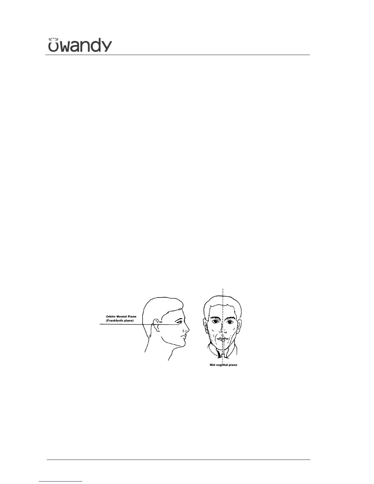
3. The patient’s head must be slightly tilted downward in order to make
the Frankfurt plane horizontal. In this way, the hard palatal ceiling
will be projected slightly over the superior apex of the anterior teeth.
If the patient has a low palatal ceiling, slightly increase the
downward tilting.
4. Align the sagittal medial plane with the centre of the chin support,
normally indicated by the relevant light beam.
Figure 18
5. The patient must extend his spine; this is normally obtained by
asking the patient to step forward, making sure that all other
conditions are unchanged. If not properly extended, the spine will
cause the appearing of a lower exposed area (clearer) in the front part
of the image.