3 Imaging programs
24 SOREDEX
206718 rev 9
3.5 Cephalometric programs
Cephalometric programs provide central projection images
of patient skull and dental anatomy. Images are utilized in
ortodontics and general diagnostics.
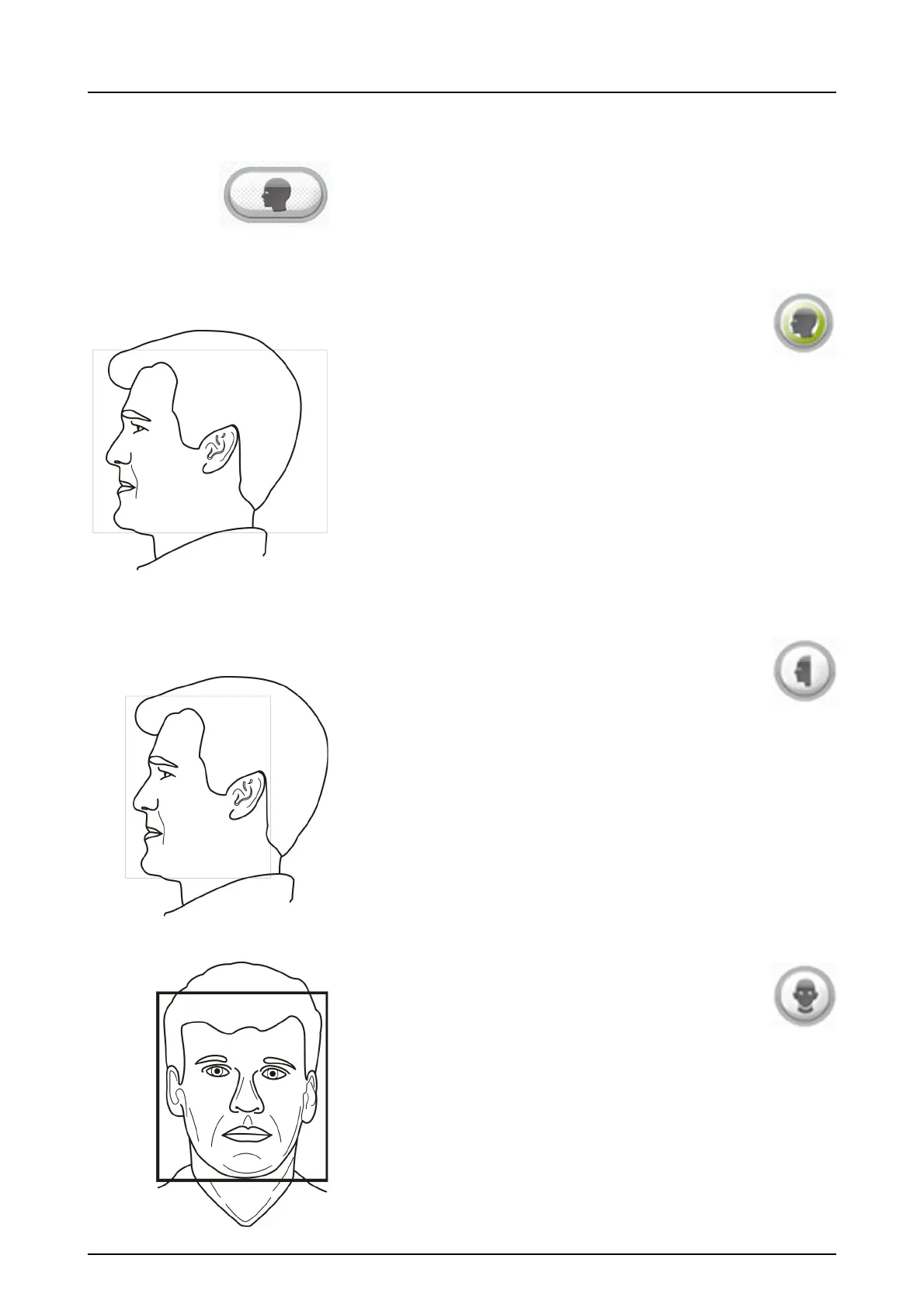
Full size lateral: Magnification 1.15
Field size: 22 x 26 cm (height x width).
Lateral Cephalostat program provides nearly full
scull image.
Reduced size lateral
Field size: 22 x 18 cm (height x width).
Reduced Width Lateral program has an optimized
image width that is used e.g. for pediatric patients but also
adult patients to reduce the radiation dose.
Cephalometric posterior-anterior (PA):
Magnification 1.15
PA 22 x 20 cm (height x width)