W
wisecandaceAug 3, 2025
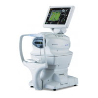
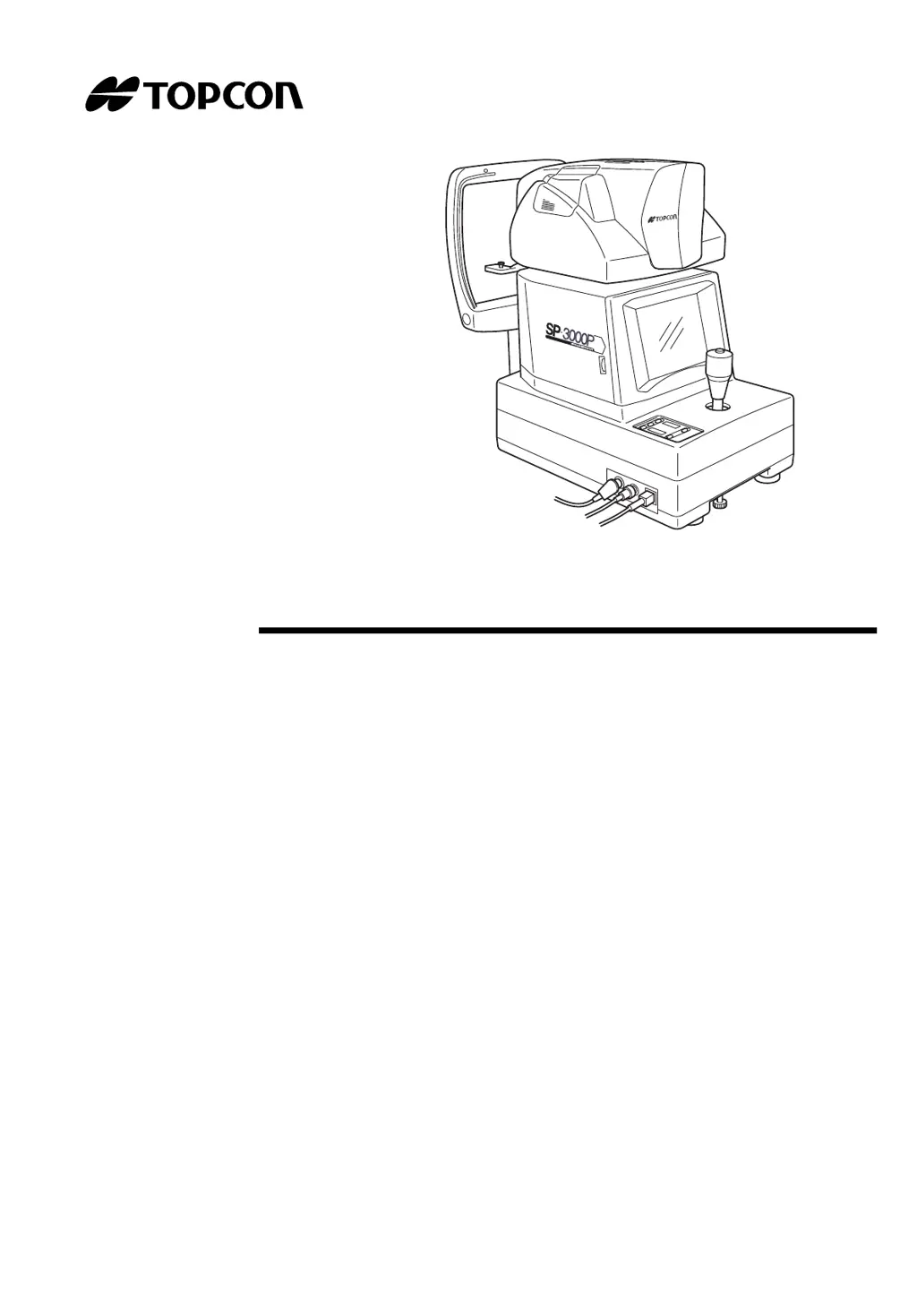
Why is nothing displayed on my Topcon SP-3000P Microscope monitor?
- JJohn TownsendAug 3, 2025
If nothing is displayed on the monitor of your Topcon Microscope, first ensure the power cord is securely plugged into the outlet and the instrument. If the issue persists, check if the Power Save condition is active and reset the instrument to normal. Finally, inspect the fuse and replace it if it has blown out.