Smartdop
®
30EX Vascular Testing Instructions
3. VENOUS COMPRESSION STUDIES
Venous compressions are auditory tests consisting of 2 maneuvers. These are, compression of the
metatarsal arch to verify augmentation followed by compression of the calf muscle to verify valvular
competence. Venous compressions are performed to detect enlarged veins due to the presence of
malfunctioning valves.
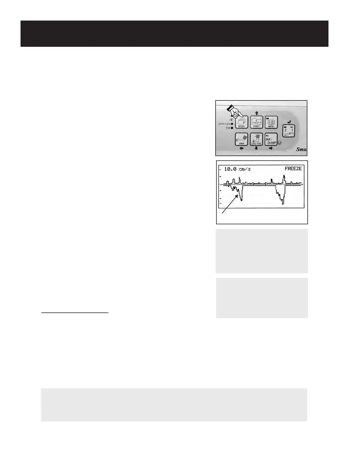
1. Connect the Doppler probe to probe connector 1 on the
right side of the unit, turn the Doppler on and press the
MODE button to select Mode 2 (venous mode) (the 2
LED will be lit).
2. Place the patient in a supine position.
3. Locate the posterior tibial artery. Then, move the probe
adjacent to the artery for diminished arterial sounds.
The probe will be in the area of the posterior tibial vein.
Listen for a “whoosh” sound.
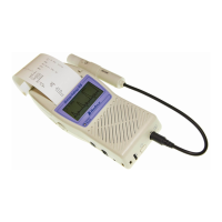
4. Hold the probe steady and rmly compress, then release
the metatarsal arch. An augmentation sound will be
heard in the form of a “whoosh” and will show below
the baseline on the LCD. This is venous blood ow
augmentation pushing venous blood past the valves
toward the vena cava.
5. Following this maneuver, continue to hold the Doppler
probe in place and squeeze the calf muscle in a down-
ward motion without moving the hand.
6. The Smartdop
®
30EX will display the dual baseline
reecting the augmented or diminished sounds. Press
the probe button or INF/DUMP button to freeze the
waveform.
Venous Augmentation
NOTE
The probe button function can
be set to FREEZE only, PRINT
only, or FRZ&PRINT. See 5-3-
15 “OTHERS-PRB-KEY” for
details.
NOTE
Patients who are hyperdynamic or who have edema may require further verication. Edema
and pregnancies may increase the risk of false positive results. Deep vein impairment requires
further testing to include duplex imaging.
Interpreting the Results
During proximal compression, a brief loud sound that rapidly concludes as blood movement is
stopped by competent valves, is expected. A long sound that continues as long as compression is
applied may indicate valvular incompetence.
As distal compression is released, blood ows backward. In competent valves, compression
release should cause a rapid termination of sound (within 1 to 5 seconds) as the competent valves
snap shut to prevent distal ow. With incompetent valves, as distal compression is released, blood
ow continues backward with a prolonged sound as incompetent valves are unable to prevent
continued ow.
57
WARNING
If any abnormality is found on
the unit or patient, discontinue
use in a manner safe to the
patient.
Weiss RA. Vascular Studies of the Legs for Venous or Arterial Disease. Dermatologic Clinics, Volume 12, Number 1, January 1994.
175-190.