Image Optimization 5-137
3. Inject the contrast agent. Enable the timer, and save the dynamic image. See also Chapter 5.13
Contrast Imaging.
5.17.9 Freehand 3D
NOTE: The effect of freehand 3D image acquisition is dependent on the technique of the
operator. Thus, there is a risk of low registration accuracy.
Operation Procedures:
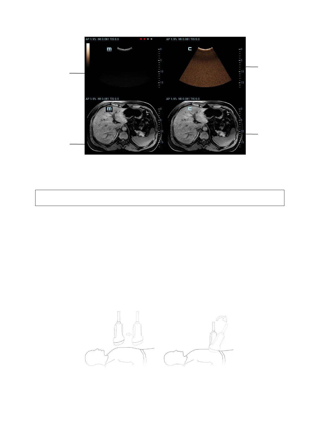
1. Select a proper probe and correct exam. Enter real-time B/Contrast mode (non-cardiac Contrast
Imaging mode).
2. Get magnetic devices prepared. Connect the magnetic devices (see also Chapter 5.17.2 Magnetic
Navigator). Power on the magnetic navigator, and check if the indicator becomes green.
3. Tap [Freehand 3D] on B-mode/Fusion-mode/Contrast-mode tab of the touchscreen.
Entering freehand 3D mode under fusion imaging mode is preferred.
4. Capture images using linear scan or rocked scan.
Linear scanning
Move the probe across the surface slowly at a constant speed.
Rocked scanning
Rotate the probe once from the left to the right side (or from the right to the left) slowly at a
constant speed to include the entire desired region.
5. After the acquisition is completed, the freehand 3D cine is automatically saved. (To set freehand
cine length, select “<F10 Setup>[System][Image]”)
If the freehand 3D cine is captured under fusion imaging mode, the system automatically registers
the ultrasound image with the freehand volume data after the acquisition is completed. Do not move
1
2
3
4
 Loading...


