5-136 Image Optimization
5. Tap [Motion Compen] to activate it. Move the probe. The Ultrasound System shows the CT image
which is processed by respiration compensation (Fusion Imaging with the respiration
compensation).
6. Save multi-frame cine.
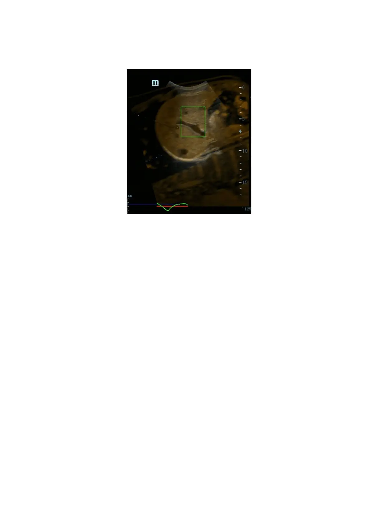
Respiration Range
The aspiration curve appears due to the active respiration depth. The respiration
curve beyond the scale becomes the straight line.
Operation Rotate the knob beneath [Resp Range].
Respiration curve scale and the unit appear on the right-axis.
5.17.8 Contrast Fusion Imaging
Contrast Fusion Imaging increases the possibility of diagnosing the difficult lesions in the pre-operation;
improves the accuracy to ablating the lesion in the intra-operation; estimates the therapeutic effect of
the target in the post-operation.
1. Tap [Contrast] to enter Contrast Fusion Imaging after the Fusion Imaging is registered.
Set fusion ratio. Adjust the display ratio of two split windows that the contrast image registers with
CT/MR/PET/freehand image. (See also Chapter 5.17.10 Parameter Setting).
2. Contrast Fusion Dual Live:
Select [Contrast][Dual Live] to adjust the fusion ratio. Adjust the display ratio that tissue image
registers with CT/MR/PET/freehand image (see Window 1 and window 2). Adjust the display ratio
that contrast image registers with CT/MR/PET/freehand image (see window 3 and window 4).
 Loading...


