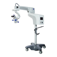
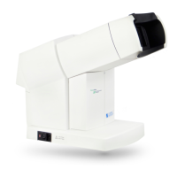
Device overview S88 / OPMI Lumera T
Version 8.0
Page 64 G-30-1682-en
Illumination system
The illumination system of the Lumera T surgical microscope contains a UV
blocking filter as a standard feature. This helps the surgeon to reduce the risk
of phototoxic retinal injury in the patient.
With SCI (Stereo Coaxial Illumination) and surrounding field illumination,
the illumination system of the surgical microscope has been specially tailored
to the requirements of ophthalmic applications.. The illumination options
provide very effective illumination of the field of view and optimum
visualization of the red reflex.
To protect the patient's eye, the illumination system is equipped with a retinal
protection device and a blue barrier filter (retina protection filter). The retinal
protection device covers the patient's pupil and prevents light from entering
the patient's eye. It is integrated in the surgical microscope and can be swung
into the beam path when the red reflex is not needed. The blue barrier filter
(retina protection filter) reduces the retinal exposure of the patient's and
surgeon's eyes and permits the radiation exposure time to be increased by
factor 3. It can be swung into the beam path of the light source in the
suspension system.
The light is supplied by a light guide which directs the light from the light
source in the suspension system to the surgical microscope.
A foot control panel is used to switch the light source on and off and to
control its brightness.
Surrounding field illumination
The surrounding field illumination is integrated in the surgical microscope and
provides an optimally illuminated field of view with superb detail recognition.
The surrounding field illumination intensity can be separately reduced or
completely deactivated using knob (1) on the surgical microscope.
Red reflex illumination
The red reflex illumination (stereo coaxial illumination) is integrated in the
surgical microscope and provides an optimally visible red reflex. For information
on how to ensure optimum red reflex visualization, please see
page 157.
 Loading...