Acclarix AX8/Acclarix AX7 Diagnostic Ultrasound System User Manual Imaging
- 35 -
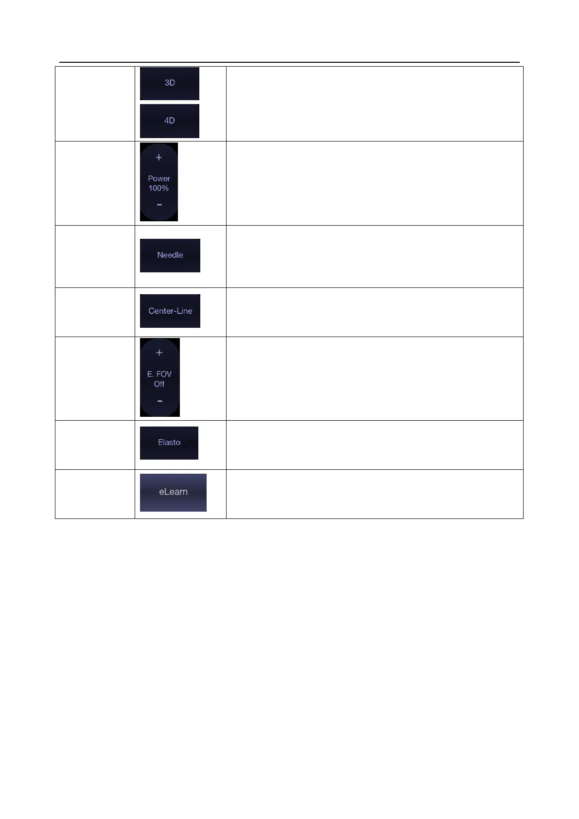
Table 5-1 B-mode Touch Screen Controls
5.2 Color Mode
5.2.1. Color Mode Variants
The system supports 3 types of Color Doppler imaging:
Color (Color Doppler): This is velocity Color Doppler that shows direction and velocity of flow.
Different colors represent different velocities and positive flow has different colors than
negative flow.
PDI (Power Doppler Imaging): PDI shows the power, or intensity, of the Doppler signal. PDI is
typically more sensitive to low levels of flow, but cannot distinguish the velocity or direction of
the flow.
DPDI (Directional Power Doppler Imaging): This is similar to DPI in that it shows the power of
the Doppler signal instead of the velocity. However, it does map positive flow to different colors
than negative flow.
Press to activate 3D/4D imaging mode. Refer to section 5.9
3D/4D mode for details.
Adjusts the acoustic output power of the activate transducer. It
is only available in live imaging. Higher acoustic power numbers
correspond to increased sensitivity in the image with improved
penetration, but the ALARA principle should be followed in
actual clinical situations.
Press to invoke the touch screen for Needle Enhancement
Visualization and Needle Biopsy Guide functions. See section
6.4 and 6.5 for more information.
Press to activate the Center Line function. Refer to section 6.6
for details.
Press to invoke extended field of view function. Only available
for linear transducers.
The extended field of view function displays as trapezoid
imaging, and its imaging width is adjustable with three levels.
Off is to disable the extended field of view function.
Press to invoke Elastography mode. Refer to section 5.11 for
details.
Press to access the instruction guide for basic scanning and for
nerve block.
 Loading...