Acclarix AX8/Acclarix AX7 Diagnostic Ultrasound System User Manual Imaging
- 47 -
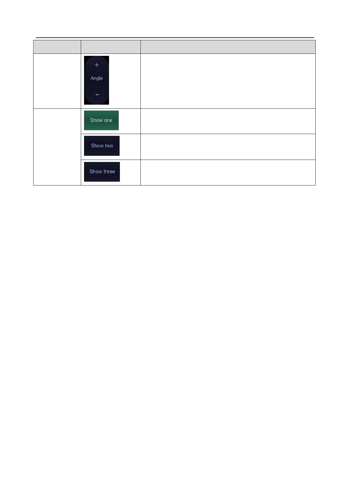
Adjusts the angle of M sample line.
Displays only one M sample line.
Displays two M sample lines.
Displays three M sample lines.
Table 5-6 Anatomic M Mode Touch Screen Controls
5.7 Color M Mode
Color M mode superimposes color encoded information on M mode strip to indicate the direction,
velocity and timing of cardiac flow and tissue movements. The direction of cardiac tissue movements
can be identified by color changes.
The Color M mode includes Color Flow M mode and Color Tissue M mode.
Note:
Color M mode is not available on transducers C5-2XQ, L10-4Q, E8-4Q (PN: 02.01.211392) and
C5-2MQ.
5.7.1. Using Color M Mode
1. Enter Color M mode
Enter Color Flow M mode
In B+M mode, press <Color>;
In B+Color mode, press <M> to display M sample line, and then press <M> again.
In B+Color+PW or B+Color+CW mode, press <M>.
Enter Color Tissue M mode
In Color Flow M mode, press TDI mode button on the touch screen.
In Color-TDI mode, press <M>.
Note:
Only phased array transducer supports Color Tissue M mode.
2. Adjust the position of ROI box or M sample line.
3. Adjust image parameters to optimize the image.
5.7.2. Color M Image Optimization
Touch screen controls that can be adjusted in Color M mode are the same as those in B mode, Color
mode and M mode, see section 5.1.2, section 5.2.3 and section 5.5.2 for details.
 Loading...