Acclarix AX8/Acclarix AX7 Diagnostic Ultrasound System User Manual Imaging
- 51 -
While scanning, the sector in the right bottom is filled gradually, and no buttons on the control
panel and touch screen can be activated.
5.9.3. 3D Image Review
The system will enter to the 3D review status after finishing one sweep.
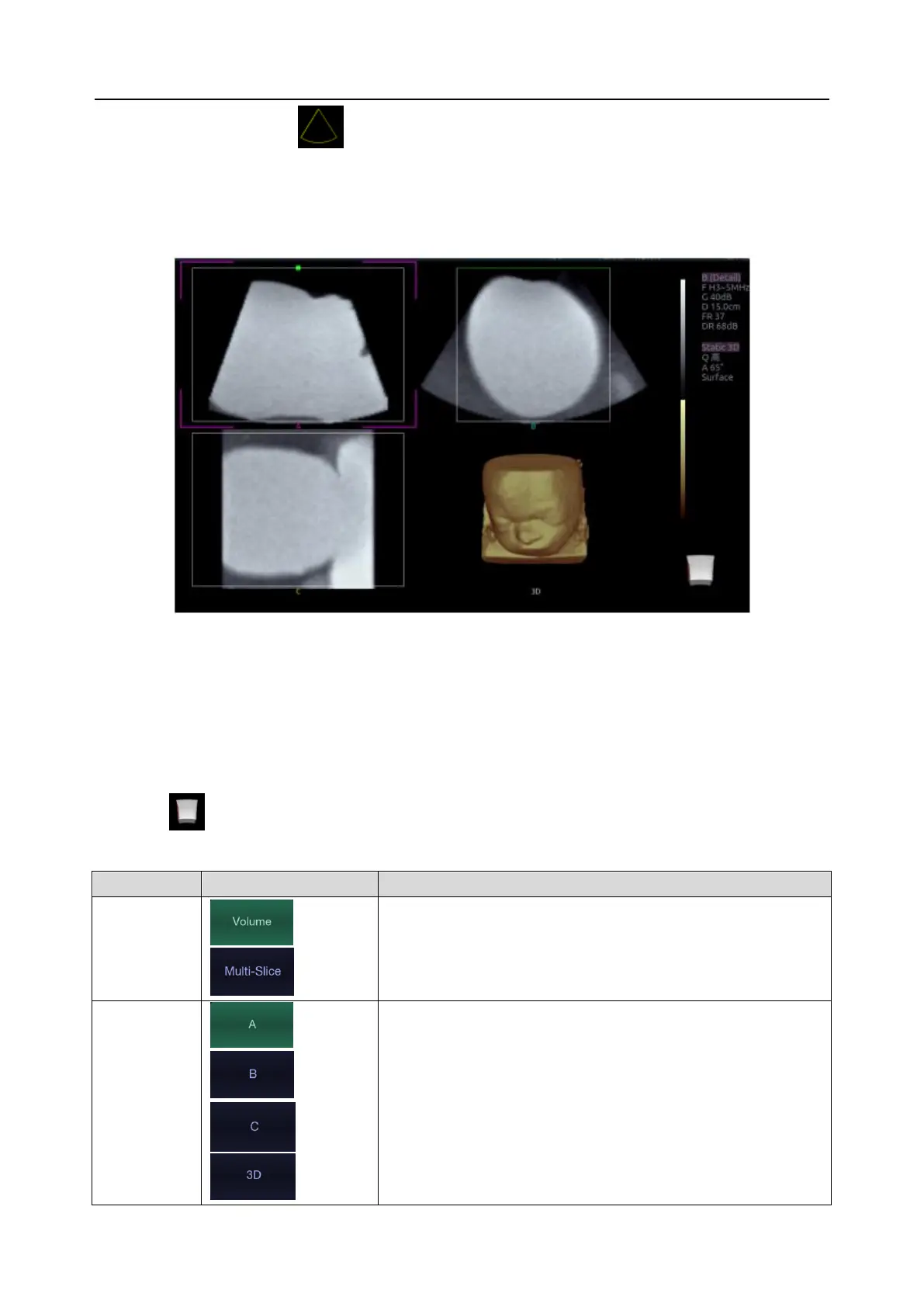
Figure 5-3 3D Image Review
There are two image modes: Volume imaging mode and multi-slice imaging mode. Figure 5-3 shows
Volume Imaging Mode in quad screen with a Baby Face volume rendering.
Quadrant A shows a slice through the data that mimics the original ultrasound image.
Quadrant B is orthogonal to A, as if the transducer had been rotated 90 degrees.
Quadrant C is orthogonal to A and B, showing a slice that is parallel to the transducer face.
The icon at the bottom-right shows the position of each slice with respect to the full 3D data set.
The following table shows the touch screen controls that are available in Volume Imaging Mode
These two radio buttons toggle between the Volume display
and the Multi-Slice display.
These four radio buttons select which quadrant is the focus
of the navigation/panning controls. A/B/C are three
orthogonal slices through the volume, while ‘3D’ is the
rendered image.
 Loading...