9 Technical data
206718 rev 9
SOREDEX 101
NOTICE! Accuracy of the imaging program factors that are
shown in GUI are:
•kV: +- 5kV
• mA: +- 1mA / +-20%
• time: +- 10%
• DAP: +- 50%
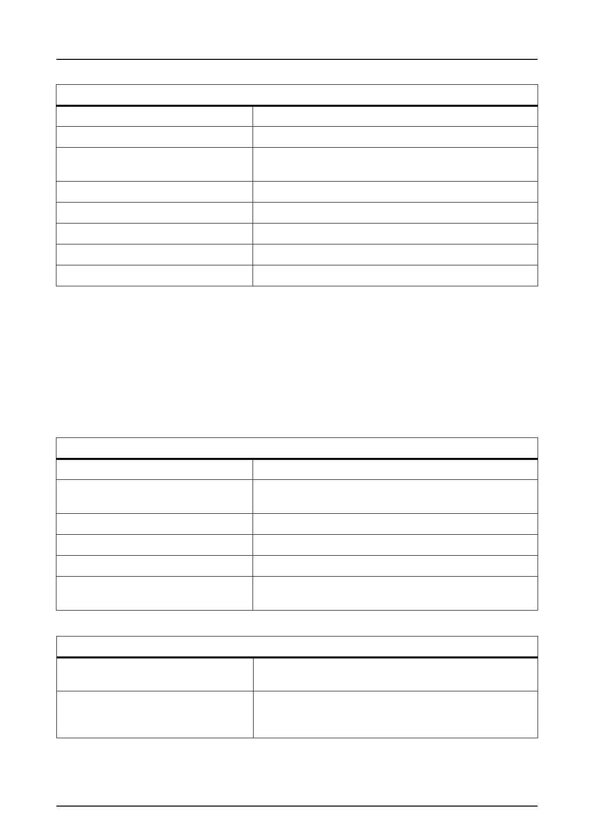
3D imaging programs:
61 x 41 mm FOV Minidose resolution 90 kV / 3.2 - 7.1 mA / 1.2 s
61 x 41 mm FOV standard resolution 90 kV / 6.3 - 12.5 mA / 2.3 s
61 x 41 mm FOV high resolution and
Endo program
90 kV / 4 - 12.5 mA / 6.1 s
61 x 41 mm FOV scout 90 kV / 4 - 12.5 mA / 0.02 s
61 x 78 mm FOV Minidose resolution 90 kV / 3.2 - 5 mA / 2.4 s
61 x 78 mm FOV standard resolution 90 kV / 6.3 - 12.5 mA / 4.9 s
61 x 78 mm FOV high resolution 90 kV / 4 - 10 mA / 12.6 s
61 x 78 mm FOV scout 90 kV / 4 - 12.5 mA / 0.04 s
Image storing and retrieving:
File formats PNG (16-bit), JPG (12-bit)
File compression PNG (lossless),
JPG (100%-60% quality)
Typical panoramic file size About 2-4 MB (PNG 16 bits)
Typical cephalometric file size 3-5 MB (PNG 16 bits)
Typical 3D file size 150-250 MB (DICOM)
Patient database Standalone workstation
Server on local area network (LAN)
Panoramic patient positioning
Operation Left
Motorised carriage movement
Positioning aids Chin rest, bite block, 3-point headrest
Hinged mirror, 3 positioning laser
lights, Y-layer step less adjustment