3-47
■
[Probe with a membrane cap attached]
<When an eye cup is not used>
4) Apply an appropriate amount of the cornea protective agent to the contact
section of the membrane cap or the examined eye.
5) Apply the tip of the membrane cap directly to the lid of the examined eye.
<When an eye cup is used>
■
Be sure to set a probe cap membrane when you use an
eye cup. The probe will be damaged if saline solution
flows into it.
6) Sink, into the eye cup, the probe to which an membrane cap is attached.
Sink the membrane cap to the saline solution keeping its brim above the
solution surface.
7) Make various settings including the total gain, the dynamic gain, the near
gain, and the far gain to obtain the best image (See “3.3.3 Adjustment by
gain setting knobs” and “3.3.7 Functions for real time mode.”)
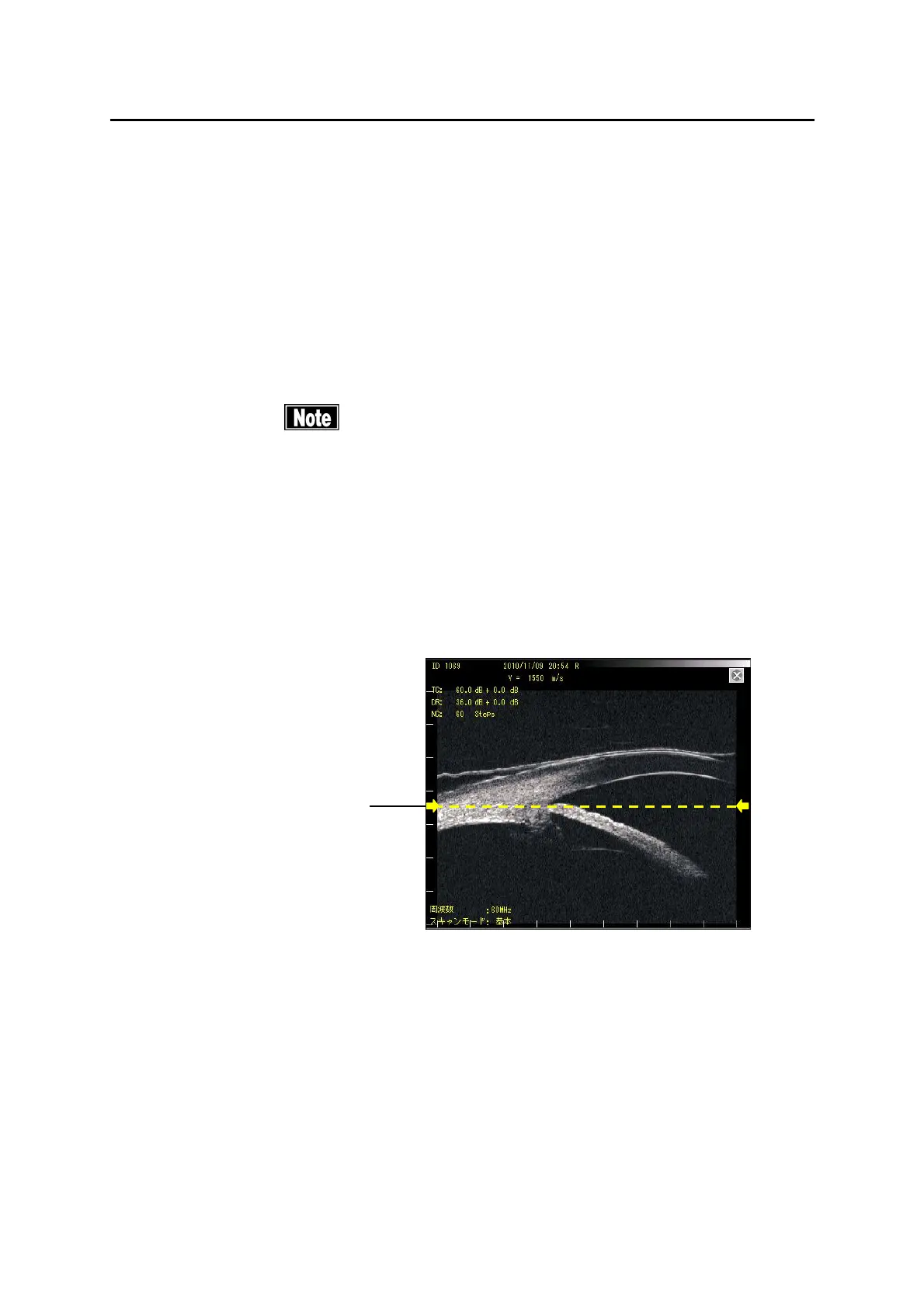
8) In the case of 60 MHz UBM probe, the best focused image is obtained in
the neighborhood of focused area marker (3) (Fig. 3). Adjust the probe
setting so that the part that you want to observe is shown in the
neighborhood of the focus area marker (3).
9) To capture the image, touch the “FREEZE” button (1) or step on the
footswitch FREEZE pedal (2) to enter the FREEZE mode.
10) To obtain another ultrasound image of the same patient, touch the “Scan”
button (1) or step on the FREEZE pedal (2) on the footswitch to release the
FREEZE mode.
(3)
(Fig. 3)
3-40
 Loading...