Ch. 6. Fluid Imaging in a Droplet Sec. 6.3. Preparing for Imaging
3.
Lower the objective:
• Lower the objective while watching for
the surface to come into focus.
• As you lower the objective, you will
first see the edge of the field diaphragm
come into focus.
• Once the field diaphragm is in focus,
slowly continue to lower the objective.
Look for subtle structures like the edge
of a layer of mica or a small bits of
debris. This is most likely the sample
surface.
4. One way to confirm this is to note the focus position distance located just below the arrow buttons.
Raise the objective back up to focus on the field diaphragm and note how much the focus distance
has changed. Typically, the sample focus distance is about 30µm below the focus distance of the
field diaphragm.
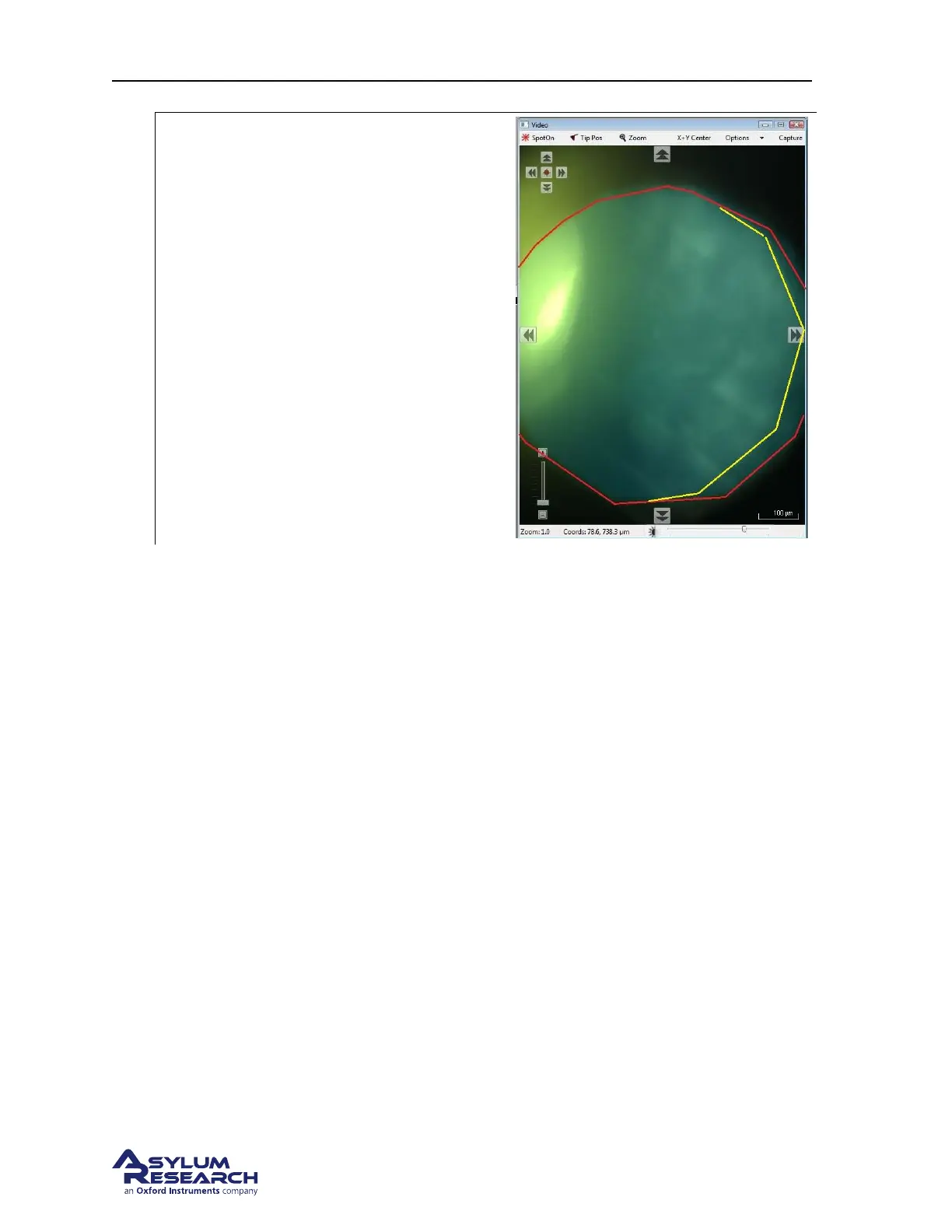
Note You may see that the edge of the field diaphragm is shifted off center. This is due to a small
amount of misalignment of the illumination path in the view module. In many cases this can help
you distinguish when the edge of the field diaphragm is in focus.
BETA
Page 64