146
3.19 Phantom Image Quality (Medical Physicist)
IMPORTANT
•••••••••••••••••••••••••••••••••••••••••••••••••••••••••••••••••••••••••••••••••••••••••••••••••••••
• The phantom image quality represents the total performance of the entire imaging chain. Changes in image quality may be
attributable to the mammography X-ray unit, REGIUS plates and cassettes, REGIUS Model 190/210/110HQ, laser printer,
lm, view box, or review workstation. Additional tests will be needed to identify the component(s), responsible for the change.
• It is also necessary to ensure that the exam tag or the processing menu has not been changed. These factors will substan-
tially aect the test results.
•••••••••••••••••••••••••••••••••••••••••••••••••••••••••••••••••••••••••••••••••••••••••••••••••••••••••••••••••••••
HINT
•••••••••••••••••••••••••••••••••••••••••••••••••••••••••••••••••••••••••••••••••••••••••••••••••••••
• Mammography phantom images should always be viewed
– By the same person
– On the same viewing device
– Under the same viewing conditions as the Radiologist review stations
– Using the same type of magnication used for reading mammograms
– Softcopy images should be window and leveled to best demonstrate the objects of interest
•••••••••••••••••••••••••••••••••••••••••••••••••••••••••••••••••••••••••••••••••••••••••••••••••••••••••••••••••••••
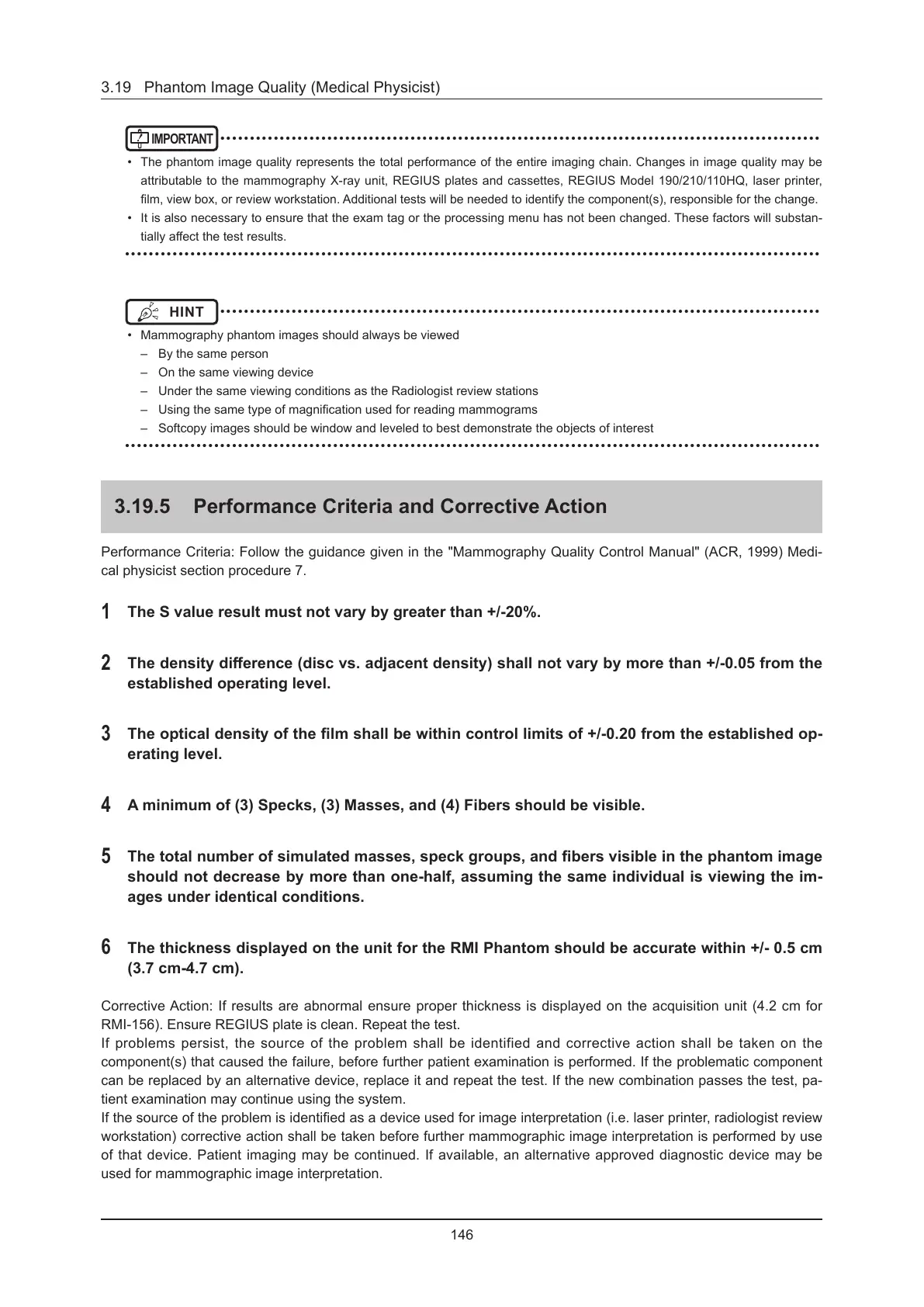
3.19.5 Performance Criteria and Corrective Action
Performance Criteria: Follow the guidance given in the "Mammography Quality Control Manual" (ACR, 1999) Medi-
cal physicist section procedure 7.
1
The S value result must not vary by greater than +/-20%.
2
The density dierence (disc vs. adjacent density) shall not vary by more than +/-0.05 from the
established operating level.
3
The optical density of the lm shall be within control limits of +/-0.20 from the established op-
erating level.
4
A minimum of (3) Specks, (3) Masses, and (4) Fibers should be visible.
5
The total number of simulated masses, speck groups, and bers visible in the phantom image
should not decrease by more than one-half, assuming the same individual is viewing the im-
ages under identical conditions.
6
The thickness displayed on the unit for the RMI Phantom should be accurate within +/- 0.5 cm
(3.7 cm-4.7 cm).
Corrective Action: If results are abnormal ensure proper thickness is displayed on the acquisition unit (4.2 cm for
RMI-156). Ensure REGIUS plate is clean. Repeat the test.
If problems persist, the source of the problem shall be identified and corrective action shall be taken on the
component(s) that caused the failure, before further patient examination is performed. If the problematic component
can be replaced by an alternative device, replace it and repeat the test. If the new combination passes the test, pa-
tient examination may continue using the system.
If the source of the problem is identied as a device used for image interpretation (i.e. laser printer, radiologist review
workstation) corrective action shall be taken before further mammographic image interpretation is performed by use
of that device. Patient imaging may be continued. If available, an alternative approved diagnostic device may be
used for mammographic image interpretation.

 Loading...