Image Optimization 5-41
5.11 3D/4D
Tips:
You can select 4D optional module. 4D module includes 4D and Static 3D
functions. You can also select Smart3D optional module only.
NOTE:
3D/4D imaging is largely environment-dependent, so the images obtained are
provided for reference only, not for confirming a diagnosis. Please compare the
images with that of other machines, or make diagnosis using none-ultrasound
methods.
5.11.1 Note before Use
5.11.1.1 3D/4D Image Quality Conditions
NOTE:
In accordance with the ALARA (As Low As Reasonably Achievable) principle,
please try to shorten the sweeping time after a good 3D imaging is obtained.
The quality of images rendered in the 3D/4D imaging is closely related to the fetal
condition, angle of a B tangent plane and scanning technique (Smart3D). The following
description uses the fetal face imaging as an example, the other parts imaging are as the
same.
Fetal Condition
Gestational age
Fetuses of 24~30 weeks old are the most appropriate for 3D imaging.
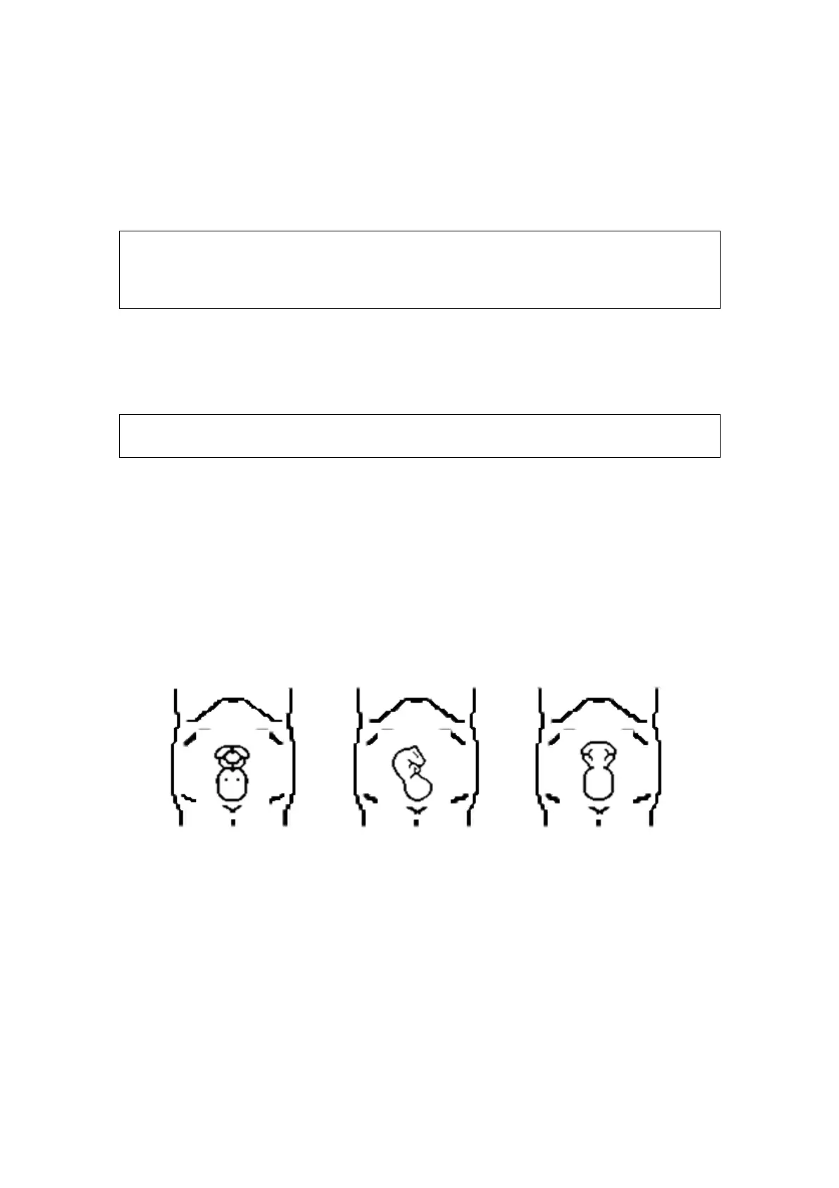
Fetal Body Posture
Recommended: Cephalic face up (figure a) or face aside (figure b).
NOT recommended: Cephalic face down (figure c).
a
b c
Amniotic fluid(AF) isolation
The region desired is isolated by amniotic fluid adequately.
The region to imaging is not covered by limbs or umbilical cord.
The best condition is that the fetus keeps still. If there is a fetal movement in
Smart 3D or Static 3D imaging, you need a rescanning when the fetus is still.
Angle of a B tangent plane
The optimum tangent plane to the fetal face 3D/4D imaging is the sagittal section of
the face, or the coronal section. To ensure high image quality, you’d better scan
maximum face area and keep edge continuity.