Image Optimization 5-43
B-section, and YZ-paralleled plane is A-section. The probe is moved along the
X-axis.
ROI (Region of Interest): a volume box used to determine the height and width of
scanning volume.
VOI (Volume of Interest): a volume box used to determine the area of a sectional
plane for 3D imaging.
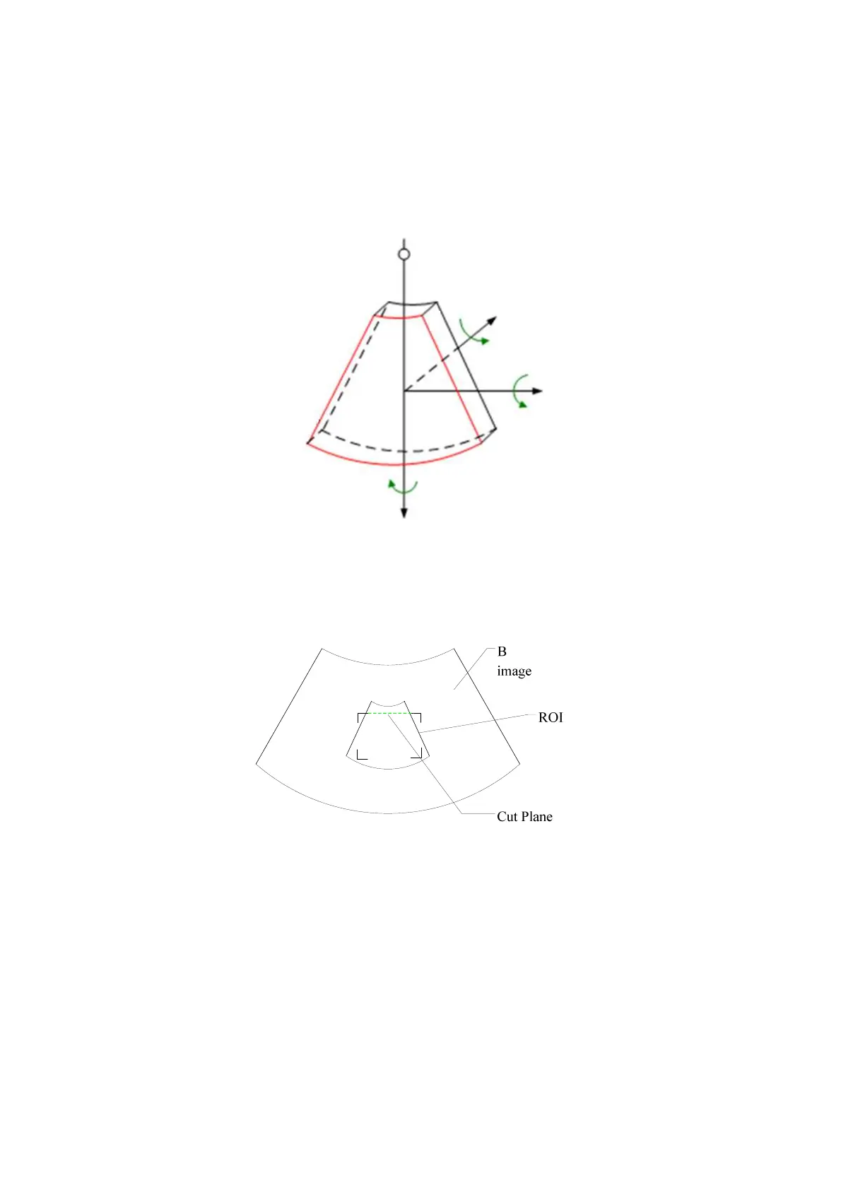
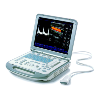
ROI and VOI
Before image acquisition after the system enters 3D/4D imaging, a B image with ROI
displays on the screen. A line (shown in the following figure) that shows the cut plane
position of VOI is inside ROI.
ROI size and position
Roll the trackball to change the ROI size and position, press the <Set> to toggle
between setting the size (dotted line) and position (solid line, with a small box at
each corner of ROI).
Curved VOI adjustment
Roll the trackball to change the curved VOI position, press <Set> to switch
among the state of changing ROI and curved VOI.
This function is to change the curved shape of the nearest VOI section, to
facilitate observation for the interested volume data.
Z
View point
X
Y
1
5
6
7
8