PK=aÉëÅêáéíáçå
fåëíêìÅíáçå=j~åì~ä=`êçëëÄÉ~ã=PQM=ÉåMOE`loF =PP=çÑ=N PQ
mêáåÅáéäÉ=çÑ=çéÉê~íáçå
3.3.2. Electron optics (GEMINI
®
column)
The GEMINI
®
column is the area of the microscope, where electrons are emitted, accelerated,
bundeled, focused, and deflected. Main characteristics of the GEMINI
®
optics are the beam boost-
er and an objective lens that consists of a combined electrostatic/electromagnetic lens doublet.
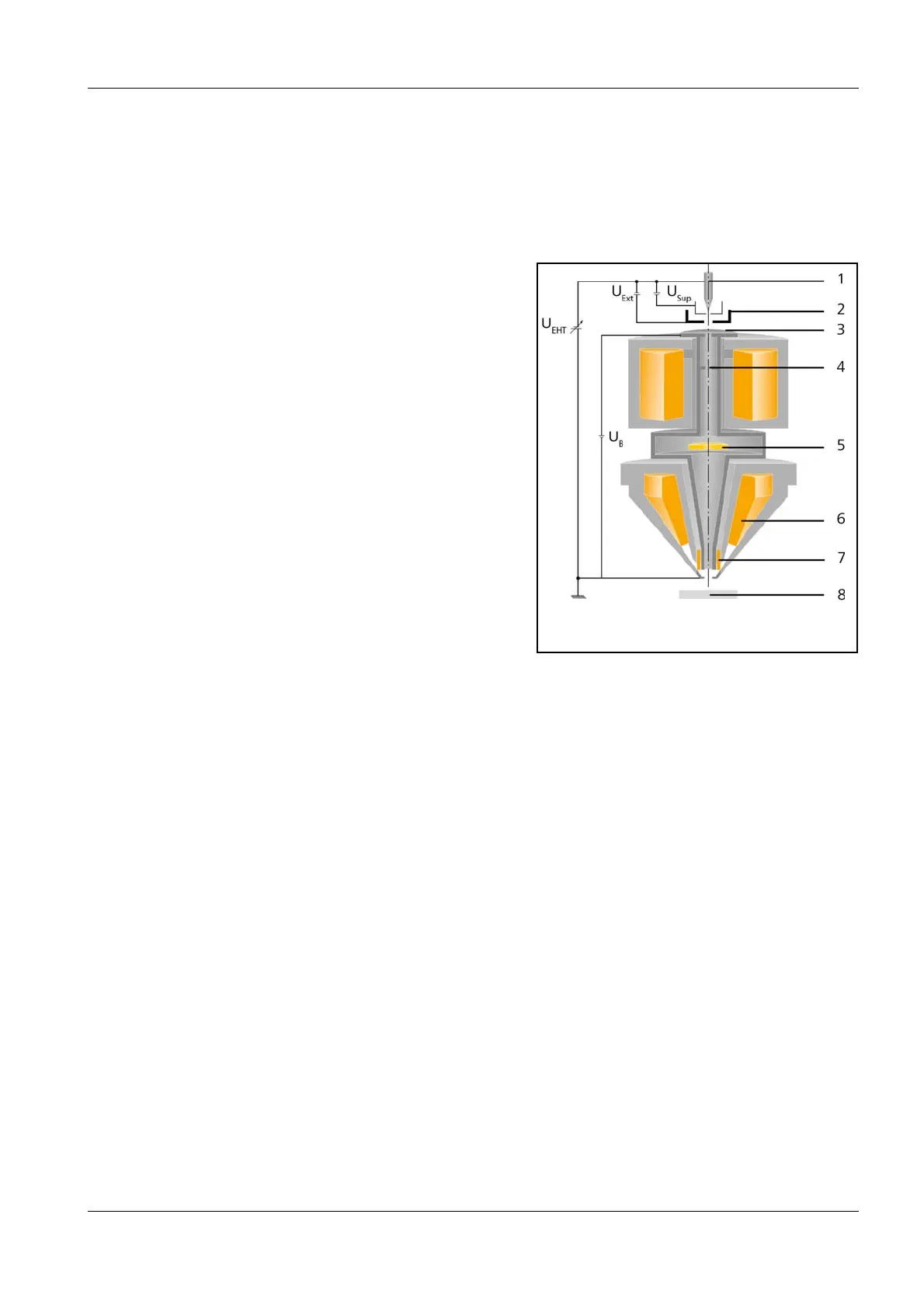
1Gun
2Extractor
3 Anode
4 Multihole aperture (aperture changer)
5 In-lens SE/In-lens Duo detector
6 Objective lens
7 Scanning coils
8 Specimen
Gun A Schottky field emitter serves as gun (1). The filament is heated by applying the filament current.
Electrons are emitted from the heated filament while an electrical field, called extractor (U
Ext
)
voltage, is applied.
To suppress unwanted thermoionic emission from the shank of the Schottky field emitter, a
suppressor voltage (U
Sup
) is applied as well.
EHT The emitted electrons are accelerated by the acceleration voltage (U
EHT
).
The beam booster (U
B
, booster voltage), which is always at 8 kV potential when the acceleration
voltage is at most 20 kV, is integrated directly after the anode. This guarantees that the energy of
the electrons in the entire beam path is always 8 kV higher than the set acceleration voltage. This
considerably reduces the sensitivity of the electron beam to magnetic stray fields and minimises
the beam widening.
Apertures The electron beam passes through the anode aperture (3) first, afterwards through the multihole
aperture (4). Standard aperture is the 30 µm aperture hole that is the central aperture. The aper-
ture size is decisive for the probe current. Other aperture sizes are selectable to meet the require-
ments of a wide range of applications. Several probe current configurations are available.
Stigmator The stigmator compensates for astigmatism, so that the electron beam becomes rotationally sym-
metrical. The electron beam is focused onto the specimen (
8) while being deflected in a point-by-
point scan over the specimen surface.
Fig. 3.7: Schematics of the electron optics