PK=aÉëÅêáéíáçå
fåëíêìÅíáçå=j~åì~ä=`êçëëÄÉ~ã=PQM=ÉåMOE`loF =PR=çÑ=N PQ
mêáåÅáéäÉ=çÑ=çéÉê~íáçå
3.3.4. Signal detection
The interaction products most frequently used for the generation of images in scanning electron
microscopy are secondary electrons (SEs) and backscattered electrons (BSEs). For the separa-
tion and detection of SEs and BSEs, for example the In-lens detector and BSD4 detector have
been developed.
FIB imaging
(optional)
FIB Electron beam is blanked,
ion beam is active. The SE sig-
nal is synchronised to the FIB
scan.
Channelling contrast imaging,
voltage contrast imaging
Defining milling patterns on the
specimen surface
Grain analysis
CrossBeam
®
operation
(optional)
SEM + FIB Image is composed of SEM
and FIB components.
Setting the coincidence point
Mill No image
Mills with the milling parame-
ters set (milling current).
Ion milling or deposition
Only deposition by ion beam
No deposition by electron beam
Mill + SEM Mills and generates a SEM
image.
SEM real-time imaging of the
ion milling or deposition
Standard detectors Configuration
Detected
signals
Typical application
In-lens SE detector
(annular SE detector)
Standard SE Surface structure
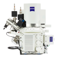
Imaging mode FIB Mode.. Characteristics Typical application
SEM imaging
FIB imaging
CrossBeam operation
Fig. 3.8: Imaging modes