5-36 Image Optimization
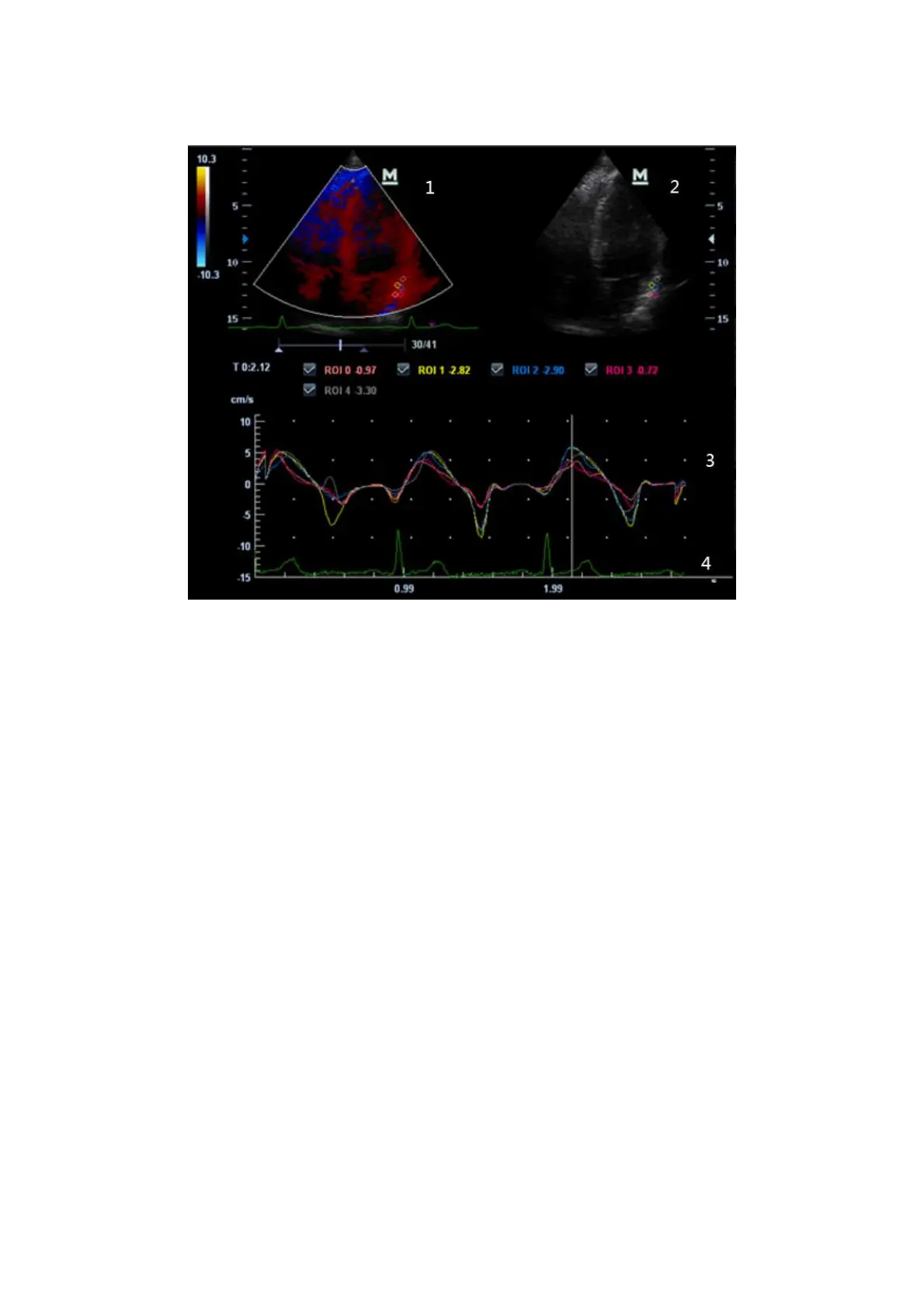
5.9.4.1 TDI QA Screen Description
1---TVI Cineloop window
Sample area: indicates sampling position of the analysis curve. The sample area is
color-coded, 8 (maximum) sample areas can be indicated.
2---B Cineloop window
Tips:
Images in the TVI cineloop window and B cineloop window are the frozen image
of the same moment; roll the trackball to review the images in the two cineloop
windows.
Sample areas are linked in the TVI cineloop window and B cineloop window.
3---Displays analysis curve
Y axis represents the velocity (unit: cm/s), while X axis represents the time (unit:
s).
Frame marker: a line that perpendicular to the X axis, can be moved horizontally
left to right (right to left) by rolling the trackball.
Click the check box beside the ROI to set if to hide or to display the TIC curve.
4---Displays ECG trace.