Do you have a question about the PerkinElmer Quantum GX2 microCT and is the answer not in the manual?
Introduces the manual, its purpose and scope.
Lists new or improved features of the Quantum GX2 microCT Imaging System.
Explains how to use tooltips for software assistance.
Lists other important documentation related to the Quantum GX2 microCT Imaging System.
Provides contact details for PerkinElmer Health Sciences.
Details how safety information is presented and defines caution/warning statements.
Shows and defines safety symbols used in the manual and on the equipment.
Provides instructions on operating the Quantum GX2 microCT safely and effectively.
Discusses X-ray safety regulations and requirements for operation and surveys.
Details environmental requirements for optimal system performance and safety.
Provides guidelines for selecting an appropriate installation environment.
Instructions for cleaning and warnings about moving the system.
Discusses power sources, voltage requirements, and power cord protection.
Mentions surge protection for electrical transient events.
Advises on actions to take during power outages.
Warns against overloading electrical outlets and equipment.
States that all servicing should be referred to PerkinElmer technical support.
Discusses the use of non-recommended equipment.
Highlights various warnings related to system operation and safety.
Warns against self-servicing and highlights high voltages behind panels.
Warns that the equipment produces X-rays when energized.
Details safety precautions related to the system's weight and moving parts.
Covers precautions for handling test samples and waste disposal.
States there are no user-serviceable components and warns about beryllium.
Discusses X-ray safety and radiation hazards.
Explains X-ray energy, shielding, and user responsibility for subject exposure.
Details X-ray dose rates and compliance with regulations.
Describes shielding, interlocks, and solenoid locks for safety.
Outlines customer responsibilities for registration and compliance with regulations.
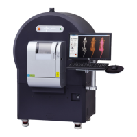
Introduces the scanner, gantry, X-ray source, and detector.
Explains the Control Panel and its functions for scan acquisition.
Details door safety features and interlocks.
Explains how to power the system and conditions for energizing the X-ray source.
Discusses the key-operated switch for controlled access and safety.
Lists specifications for the scanner and the acquisition computer.
Details specifications for the computer system.
Provides specifications for the computer monitor.
Lists environmental requirements for the system.
Describes the tube inlet for external devices and accessories.
Explains the use of the transfer bed and Mouse Imaging Shuttle for subject transport.
Details the FMT mouse imaging cassette for subject transport and registration.
Presents an overview of acquiring CT data on the system.
Details the steps for acquiring CT data on the Quantum GX2 microCT.
Describes the procedure for starting the Quantum GX2 microCT Imaging System.
Provides step-by-step instructions for powering on and launching the software.
Explains how to create a new database for image data.
Details how to connect to an existing database.
Explains how image data is organized within a database.
Covers importing, exporting, moving, and deleting data.
Discusses copying, searching, and deleting databases.
Explains how to log in and out as a system administrator.
Discusses using software for offline data analysis.
Details the steps for creating a new database in the Database window.
Provides instructions for connecting to an existing database.
Explains how samples, studies, and series are associated in a database.
Describes how to hide or show columns in the Database window.
Covers importing and exporting reconstructed image data.
Steps for importing *.vox data into the database.
Details the process for exporting image data in various formats.
Instructions on how to move a series to a different sample and study.
Explains how to delete data and the implications.
Covers copying, searching, and deleting databases.
Provides steps to copy a database for data sharing.
Details how to search for specific data within databases.
Explains how to delete a database and its associated data.
Instructions for administrator login and logout.
Introduces software for offline data viewing and analysis.
Lists requirements for offline analysis workstations.
Provides steps for setting up an analysis workstation.
Explains the warm-up procedure for the imaging system.
Guides on selecting a database and location for saving image data.
Details how to automatically export DICOM images after scans.
Explains how to set scan parameters using presets or custom settings.
Guides on selecting preset scan conditions.
Instructions for preparing and placing a subject in the sample chamber.
Step-by-step guide for performing a CT scan without gating.
Instructions for performing a CT scan with respiratory gating.
Steps for performing a CT scan with cardiac gating.
Guides on reconstructing subvolumes or slices from scan data.
Detailed steps for reconstructing subvolumes.
Detailed steps for reconstructing slices.
Details acquiring images in fluoroscopy mode.
Explains the ring reduction algorithm and how to use it.
Describes how to change X-ray filters to optimize protocols.
Explains how the Database window provides previews of 3D reconstructions.
Describes the AutoViewer for viewing scans and reconstructions.
Details the Viewer for 2D image display and analysis.
Explains how to view Z-axis slices and adjust viewing options.
Details the 3D Viewer for rendering and visualizing 3D reconstructions.
Explains how to classify data using the 3D Viewer.
How to edit color tables and manage classified data parts.
How to adjust detail level and shading in the 3D Viewer.
Recommends periodic X-ray leakage safety tests.
Explains the complexity and need for expert radiation surveys.
Details daily, weekly, monthly, and annual safety checks.
Lists daily and weekly safety checks to perform.
Outlines monthly safety checks, including the emergency off switch.
Lists annual safety checks, including a full radiation survey.
Discusses approved cleaning solutions and procedures.
Lists acceptable cleaning solutions for the sample bore.
Introduces the manual and PerkinElmer.
Details the limited warranty for the system and software.
Contains disclaimers regarding software and system use.
Disclaimers related to software usage and intellectual property.
Disclaimers regarding system operation and user training.
Lists issues and corrective actions for troubleshooting.
Lists user-replaceable parts and their part numbers.
| Category | Micro-CT Imaging System |
|---|---|
| X-ray Source | Microfocus X-ray tube |
| Detector | Flat panel detector |
| Maximum Field of View | 90 mm diameter |
| Scan Time | Application Dependent |