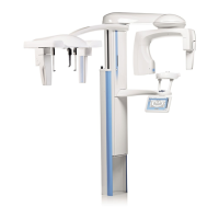
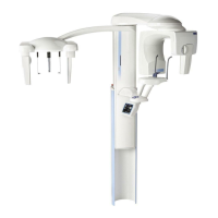
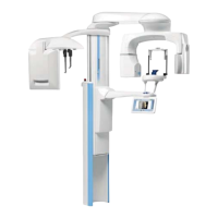
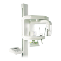
Planmeca ProMax 3D Max with ProTouch 1
TABLE OF CONTENTS
User’s Manual
1 INTRODUCTION ..............................................................................................1
2 ASSOCIATED DOCUMENTATION .................................................................2
3 SYMBOLS ON PRODUCT LABELS ................................................................3
4 SAFETY PRECAUTIONS ................................................................................3
5 SWITCHING X-RAY UNIT ON .........................................................................6
6 MAIN PARTS ...................................................................................................7
6.1 General view of X-ray system ........................................................................................ 7
6.2 General view of X-ray unit .............................................................................................. 8
6.3 Patient supports ............................................................................................................. 9
6.4 Exposure switch ........................................................................................................... 10
6.5 Emergency stop button ................................................................................................ 11
6.6 Touch screen ............................................................................................................... 12
6.7 Patient positioning controls .......................................................................................... 14
7 PROGRAMS ...................................................................................................16
7.1 3D Dental ..................................................................................................................... 16
7.2 3D Ear Nose Throat ..................................................................................................... 17
7.3 3D Models .................................................................................................................... 17
8 3D PATIENT EXPOSURE ..............................................................................18
8.1 Preparing X-ray system ............................................................................................... 18
8.2 Selecting exposure settings ......................................................................................... 21
8.3 Preparing patient .......................................................................................................... 27
8.4 Patient positioning ........................................................................................................ 27
8.5 Adjusting volume position ............................................................................................ 29
8.6 Taking a scout image or 2D views (LAT, PA or LAT-PA) ............................................ 33
8.7 Taking a 3D exposure .................................................................................................. 35
9 3D FACE PHOTO ...........................................................................................37
9.1 Before exposure ........................................................................................................... 37
9.2 Selecting exposure settings ......................................................................................... 37
9.3 Patient positioning ........................................................................................................ 37
9.4 Taking a 3D face photo ................................................................................................ 39
10 3D MODEL EXPOSURE ................................................................................40
10.1 Calibrating X-ray unit for impression or plaster material .............................................. 40
10.2 Taking an exposure of an impression or plaster cast .................................................. 44
11 SETTINGS ......................................................................................................47
11.1 User settings ................................................................................................................ 47
12 CLEANING .....................................................................................................50
13 SERVICE ........................................................................................................50
14 DISPOSAL .....................................................................................................51
15 HELP MESSAGES .........................................................................................52
16 ERROR MESSAGES .....................................................................................54