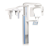
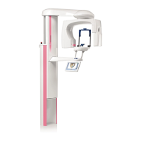
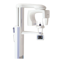
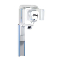
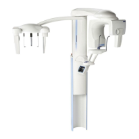
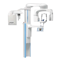
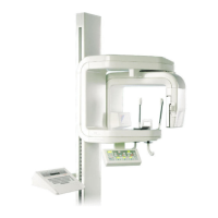
Planmeca ProMax 3D Max with ProTouch 1
INTRODUCTION
User’s Manual
1 INTRODUCTION
This manual describes how to take 3D exposures. The
manual applies to:
• Planmeca ProMax 3D Max X-ray unit
NOTE This manual is valid for software version 3.2.0.0.r or
later. This software version is compatible with
Planmeca Romexis software version 3.3.1.r or later.
To check the software version of your X-ray unit, select
Service spanner > About > 4100 Component
Information > ProMax SW version.
The X-ray unit uses cone beam computed tomography to
produce three-dimensional X-ray images. If the X-ray unit
has a ProFace sensor, you can take a 3D photo of the
patient’s face.
The X-ray images can be used to examine:
• Dentomaxillofacial area
• Regions around ear, nose and throat
• Other cranial anatomies
The 3D face photo can be used for patient education or in
order to follow the results of medical treatments.
You need a PC with the Planmeca Romexis program in
order to save, view and modify the images.
Make sure that you are fully acquainted with the
appropriate radiation protection measures and these
instructions before you use the X-ray unit.
NOTE The X-ray unit may be used by health care
professionals only.
Service
(Top left
corner of
main view)
spanner