MM098-002 – LOGOS ONE – Operator Manual
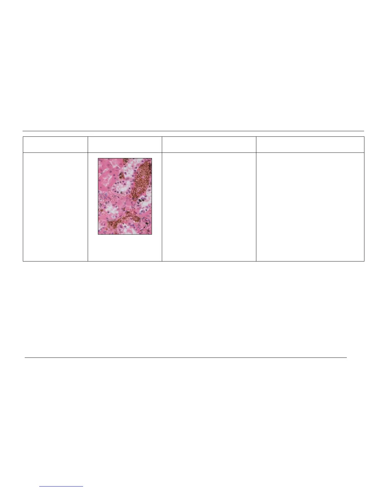
Pigment deposits in
microscopic section.
Microscopic picture shows
pigment deposits
(black/brown) at sites of
hemorrhagic deposits (Blood
vessels, Spleen and products
of conception).
1. Hemorrhagic tissue that has not been
adequately fixed (stabilized) will display
deposits of formalin pigment at sites
where there are abundant red blood
cells.
Present in tissues with hemorrhagic sites
such as major blood vessels, where there
are abundant red blood cells and related
breakdown deposits, this especially includes
the Spleen.
1. Confirm that the pigment is actually formalin
pigment and not hemosiderin (Iron break
down products of red blood).
AND
To remove pigment retrospectively, if it
interferes with diagnosis, then use the
standard formalin pigment removal treatment
method that uses Picric Acid.
AND
Prospectively, to avoid this from occurring is
to ensure that hemorrhagic tissue is well fixed
before processing.