AU-OPR-AureFloFT-EN,
Rev H
43
Transit-Time Ultrasound
A Transonic
®
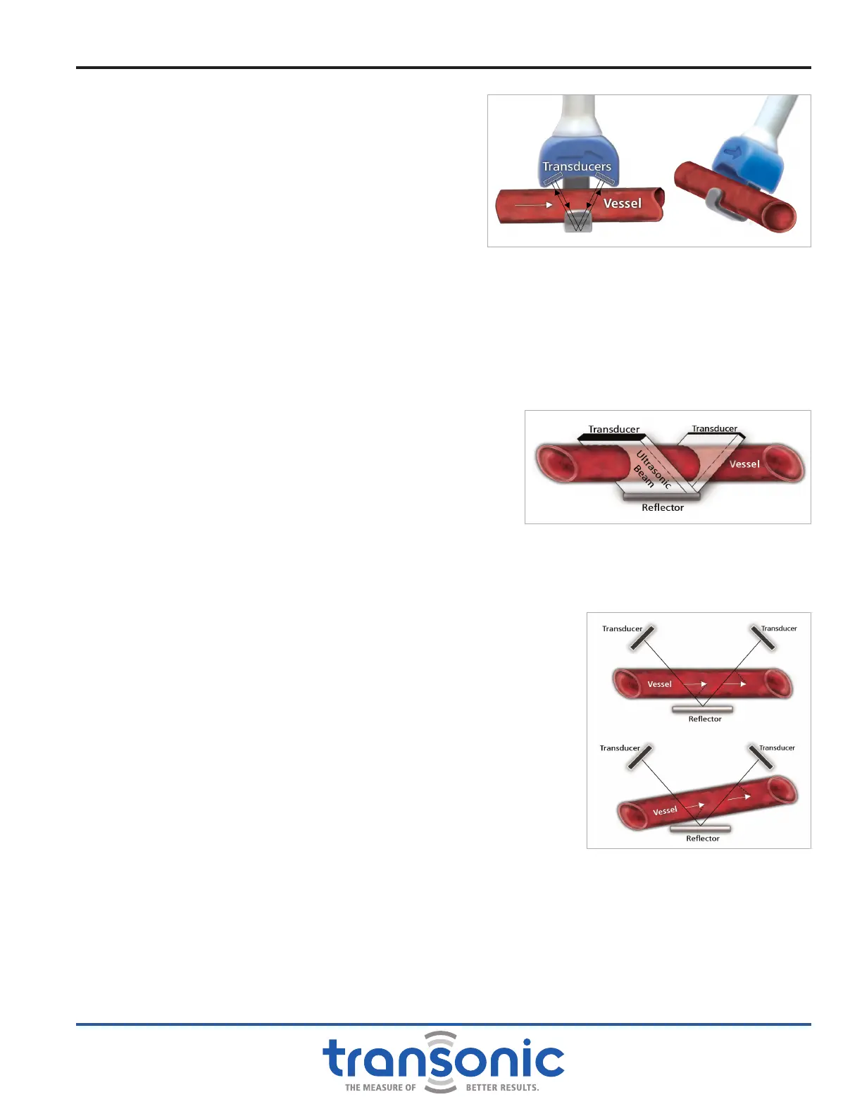
Perivascular Flowprobe (Fig. E.1) consists of a
Probe body which houses ultrasonic transducers and a xed
acoustic reector. The transducers are positioned on one
side of the vessel or tube under study and the reector is
positioned at a xed position between the two transducers
on the opposite side. Electronic ultrasonic circuitry directs a
Flowprobe through the following cycles:
UPSTREAM TRANSIT-TIME MEASUREMENT CYCLE
An electrical excitation causes the downstream transducer
to emit a plane wave of ultrasound. This ultrasonic wave
intersects the vessel or tubing under study in the upstream
direction, then bounces off the xed "acoustic reector." It
again intersects the vessel and is received by the upstream
transducer where it is converted into electrical signals. From
these signals, the Flowmeter derives an accurate measure
of the "transit time" it takes for the wave of ultrasound to
travel from one transducer to the other.
DOWNSTREAM TRANSIT-TIME MEASUREMENT CYCLE
The same transmit-receive sequence is repeated, but with the
transmitting and receiving functions of the transducers reversed
so that the ow under study is bisected by an ultrasonic wave
in the downstream direction. The Flowmeter again derives and
records from this transmit-receive sequence an accurate measure of
transit time it takes for the wave of ultrasound to travel from one
transducer to the other.
Just as the speed of a swimmer depends, in part, on water
currents, the transit time of ultrasound passing through a conduit
is affected by the motion of liquid owing through that vessel.
During the upstream cycle, the sound wave travels against ow and
total transit time is increased by a ow-dependent amount. During the
downstream cycle, the sound wave travels with the ow and total transit
time is decreased by the same ow-dependent amount. Using wide beam
ultrasonic illumination, the Flowmeter subtracts the downstream transit
times from the upstream transit times. This difference in the integrated
transit times is a measure of true volume ow.
WIDE BEAM ILLUMINATION
One ray of the ultrasonic beam undergoes a phase shift in transit time
proportional to the average velocity of the liquid times the path length over
which this velocity is encountered. With wide-beam ultrasonic illumination
(Fig. E.2), the receiving transducer integrates these velocity-chord
products over the vessel's full width and yields volume ow: average
velocity times the vessel's cross sectional area. Since the transit time
is sampled at all points across the vessel diameter, volume ow
measurement is independent of the ow velocity prole. Ultrasonic
beams which cross the acoustic window without intersecting the
vessel do not contribute to the volume ow integral. Volume ow
is therefore sensed by perivascular Probes even when the vessel is
smaller than the acoustic window (Fig. E.3).
Appendix E: Theory of Operation
Fig. E.1: Schematic views of a Transonic
®
perivascular
ultrasonic volume Flowsensor. Using wide beam
illumination, two transducers pass ultrasonic signals back
and forth, alternately intersecting the owing liquid in
upstream and downstream directions. The Flowmeter
derives an accurate measure of the “transit time” it takes
for the wave of ultrasound to travel from one transducer
to the other. The difference between the upstream and
downstream integrated transit times is a measure of
volume ow rather than velocity.
Fig. E.2: The vessel is placed within a beam that
fully and evenly illuminates the entire blood vessel.
The transit time of the wide beam then becomes a
function of the volume ow intersecting the beam,
independent of vessel dimensions.
Fig. E.3: The ultrasonic beam intersects the vessel
twice on its reective path. With each intersection,
the transit time
through the vessel is modied by a
vector component of ow. The full transit time of the
ultrasonic beam senses the sum of these two vector
components, or ow. With misalignment (bottom),
one vector component of ow increases as the other
decreases, with little consequence to their sum.
Drost, C.J., "Vessel Diameter-Independent Volume Flow Measurements Using Ultrasound", Proceedings San Diego Biomedical
Symposium, 17, p.299-302, 1978. U.S. PATENT 4,227,407, 1980.