88 BD LSR II User’s Guide
Intracellular Calcium Concentration
Flow cytometry can be used to measure the concentration of intracellular free
calcium ions. Measurement of calcium ion (Ca
++
) concentration can be made on
large numbers of single cells, which provides information about the number of
responding cells as well as the relative magnitude of the response to a given
stimulus. Ca
++
concentration can be correlated with other parameters, such as
time, phenotype, and cell cycle.
In their resting state, eukaryotic cells maintain an internal Ca
++
concentration far
less than that of the extracellular environment. Elevation in intracellular Ca
++
concentration is often used as an indicator of cellular activation in response to a
stimulus. Calcium flux is also an indicator of whether the cells in a population
remain functional after exposure to a drug or other compound.
Several fluorescent dyes measure intracellular Ca
++
levels. For most of them, the
amount of Ca
++
entering a cell is indicated by a change in fluorescence emission.
For example, the emission spectrum of indo-1 changes from blue to violet upon
binding to Ca
++
. The ratio of violet to blue fluorescence is independent of the
amount of dye within the cell.
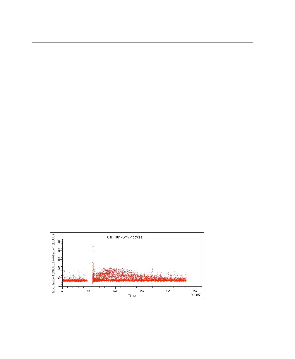
When normal cells are analyzed for calcium flux with indo-1 by flow cytometry,
a shift in the violet/blue ratio is obtained (Figure 5-1). A break in data occurs
when the stimulus is added to the sample tube. The increase in the ratio over time
reflects the increase in intracellular Ca
++
concentration.
Figure 5-1 Calcium flux data
LSR2.book Page 88 Tuesday, April 25, 2006 3:34 PM