OPERATION
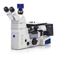
Axio Vert.A1
Illumination and Contrast Techniques in Reflected Light Carl Zeiss
05/2012 431030-7044-001 87
4.12 Illumination and Contrast Techniques in Reflected Light
4.12.1 Reflected Light Bright Field
Reflected light bright field microscopy is the simplest and most common optical microscopy technique
designed to examine opaque specimens or objects, such as polished material faces or wafers.
The light coming from and concentrated by the reflected light illuminator is reflected on a neutral-colored
beam splitter and subsequently passes through the objective, which focuses the beams on the surface of
the specimen (also known as condenser function). The objective collects the light reflected off the object
and, together with the tube lens, generates the microscopic intermediate image, which can subsequently
be observed visually or documented objectively.
Requirements
− The microscope must have properly been put into operation, as described in Section 3.
− The microscope must be switched on.
− VIS-LED or HAL 100 reflected light illumination
Setting Bright Field
• Place the specimen on the specimen stage (Fig. 4-14/1) and focus at weak magnification, e.g., using
the EC Epiplan 10x objective and the focusing drive (Fig. 4-14/3). Set the position for bright field on
the reflector turret (Fig. 4-14/2).
• Close the luminous field diaphragm (Fig. 4-14/7) until its edge becomes visible in the field of view. Use
ball-headed screwdriver SW 3 to center the luminous field diaphragm to the edge of the field of view.
• Close the aperture diaphragm (Fig. 4-14/6) slightly. Remove the eyepiece.
• Use the centering screws to center the aperture diaphragm.
• Use the adjustment wheel to set the size of the aperture diaphragm for specimens with medium-level
contrast properties to approximately two thirds to four fifths of the objective exit pupil diameter.
Reinsert the eyepiece.
The aperture diaphragm is not meant to adjust image brightness.