Faxitron CT Specimen Radiography System User Guide
Chapter 1: Introduction
5081-9544 Revision 004 Page 1
1: Introduction
Read all this information carefully before operating the system. Follow all warnings and
precautions as stated in this manual. Keep this manual available during procedures.
Physicians should tell patients about all potential risks and adverse events described in
this manual with respect to the operation of the system.
Note
Hologic configures some systems to meet specific requirements. Your system
configuration may not have all the options and accessories included in this manual.
1.1 Intended Use
United States federal law restricts this device to use by, or on the order of, a physician.
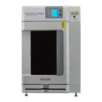
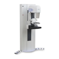
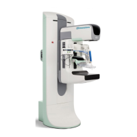
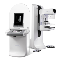
The Faxitron
®
CT is a Cabinet x-ray system that is used to provide two and three-
dimensional digital x-ray images of harvested specimens from various anatomical
regions in order to provide rapid verification that the correct tissue has been excised
during the biopsy procedure.
Doing the verification directly in the same room or nearby enables cases to be completed
faster, thus limiting the time the patient needs to be under examination. Specimen
radiography can potentially limit the number of patient recalls.
1.2 System Capabilities
The Faxitron CT system acquires and displays 2D and 3D radiographic images of
surgical specimens taken from various anatomical regions. The system has the capability
to transfer the images to external devices. The images acquired with this system are
intended to confirm removal of a suspected lesion or pathology; the system is not
intended for diagnostic purposes.
Specimen radiography systems are used in diagnostic imaging departments, pathology
departments, or in surgical suites. Radiologic Technologists, surgical personnel,
surgeons, radiologists, and pathologists can use specimen radiography equipment.
Hologic recommends that users receive training in basic ionizing radiation safety before
using the system.