Faxitron CT Specimen Radiography System User Guide
Chapter 6: Faxitron CT Software Operation
5081-9544 Revision 004 Page 71
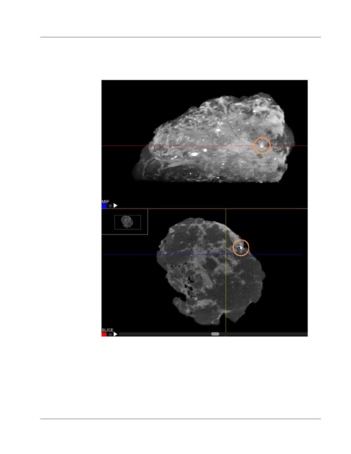
calcifications can be seen relative to the true edge of the specimen in the corresponding
view below.
Figure 88: Faxitron CT MIP and Slice Views
To switch between MIP and SLICE view simply click on the word in the bottom left
corner.
6.8.4 Hounsfield Units
The X, Y, and Z position of the cursor on the specimen, plus the Hounsfield unit, circled,
are displayed in the top right of the view window.