91
SPECIFICATIONS AND PERFORMANCE
SYSTEM CLASSIFICATION
• Types of protection against electric shocks:
This instrument is classified as Class I equipment.
Class I equipment does not depend only on basic insulation for protection
against electric shocks, but also provides a means of connection to a protec-
tive earth system of facilities so that metal parts that come into contact do not
become conductive while the basic insulation is in failure.
• Grade of protection against electric shocks:
This instrument is classified as Type B equipment.
Type B equipment provides a specified grade of protection to prevent electric
shocks, particularly for reliability against current leaks, measuring current and
protectiove earth current (in case of Class Iepuipment).
• Degree of protection against harmful ingress of water: IPx0
TRC-NW200 has no protection against ingress of water. (The degree of pro-
tection against harmful ingress of water defined in IEC 60529 is IPx0.)
• Classification according to the method(s) of sterilization or disinfection recom-
mended by the manufacturer: not applicable.
TRC-NW200 has no part to be sterilized or be disinfected.
• Classification according to the degree of safety of application in the presence of
a flammable anesthetic mixture with air or with oxygen or nitrous oxide: Equip-
ment not suitable for use in the presence of a flammable anesthetic mixture
with air or with oxygen or nitrous oxide.
TRC-NW200 should be used in environments where no flammable anesthet-
ics and/or flammable gases are present.
• Classification according to the mode of operation: Continuous operation.
Continuous operation is the operation under normal load for an unlimited
period, without the specified limits of temperature being exceeded.
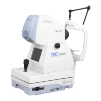
DIMENSIONS AND WEIGHT
Dimensions : 272mm (W) × 505mm (D) × 530 ~ 560mm (H)
Weight : 24kg
PURPOSE OF USE
Used to observe and photograph the retinal oculi.
OPERATION PRINCIPLE
The infrared light is emitted from the observation illumination optical system. By
using this infrared light, the eye ground of the patient is illuminated. The image
received by the built-in observation CCD camera is shown on the monitor and the
inspector can observe the eye ground. After adjusting the photography position
and focus, a visible light is emitted from the photography illumination optical sys-
tem by operating the photography switch of the instrument body. The image
received by the photography CCD camera is recorded in the memory card, a
commercial computer and an external recording device. When digital zoom is
set, the illumination aperture is automatically changed and the photographed cor-
neal diameter is changed. While the infrared filter is removed from the observa-
tion illumination optical system, color photography is possible.
 Loading...