GE Voluson E10 Training Manual
© 2017 Conquest Imaging
Module 3 Operating Modes
The following general operating modes are available on Voluson E10
systems:
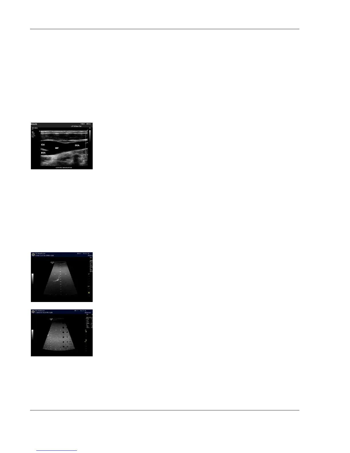
B-Mode
B-Mode is a two-dimensional image of the amplitude of the echo
signal. It is used for location and measurement of anatomical
structures and for spatial orientation during operation of other
modes. In B mode, a two-dimensional cross-section of a three-
dimensional soft tissue structure such as the heart is displayed in real
time.
Ultrasound echoes of different intensities are mapped to different
gray scale or color values in the display. The outline of the 2D (B-
Mode) (B-Mode) cross-section is a sector, depending on the particular
transducer used. B-mode can be used in combination with any other
mode.
Harmonic Imaging
Tissue Harmonic Imaging, acoustic aberrations due to tissue, are
minimized by receiving and processing the second harmonic signal
that is generated within the insonified tissue. Coded Harmonics
enhances near field resolution for improved small parts imaging as
well as far field penetration. It diminishes low frequency amplitude
noise and improves imaging technically difficult patients.
It may be especially beneficial when imaging isoechoic lesions in
shallow-depth anatomy in the breast, liver and hard-to-visualize fetal
anatomy. Coded Harmonics may improve the B-Mode (2D (B-Mode))
image quality without introducing a contrast agent.