GE Voluson E10 Training Manual
© 2017 Conquest Imaging
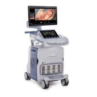
M-Mode
In M-mode, soft tissue structure is shown as a scrolling display, with
depth on the Y-axis and time on the X-axis. It is mostly used for
cardiac measurements. M-mode is also known as T-M mode or Time-
Motion mode. Ultrasound echoes of different intensities are mapped
to different gray scale values in the display. M-mode displays time
motion information derived from a stationary beam. M-mode is
normally used in conjunction with a 2D (B-Mode) (B-Mode) image for
spatial reference.
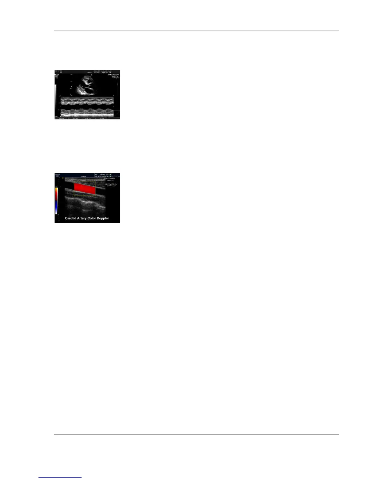
Color Flow Doppler Mode
Color Doppler is used to detect motion presented as a two-
dimensional display. There are three applications of this technique:
Color Flow Mode - used to visualize blood flow velocity and
direction.
Power Doppler (Angio) - used to visualize the spatial distribution
of blood.
Tissue Velocity Imaging - The Tissue Color Doppler Imaging is
used for color encoded evaluation of heart movements. Tissue
Velocity Imaging images provide information about tissue motion
direction and velocity.
Blood flow is displayed as a real-time two-dimensional cross-sectional
image. The 2D (B-Mode) (B-Mode) cross-section is presented as a full
color display, with various colors being used to represent blood flow
(velocity, variance, power and/or direction).
To provide spatial orientation, the full color blood flow crosssection is
overlaid on top of the gray scale cross-section of soft tissue structure
(2D (B-Mode) (B-Mode) echo). Blood velocity is the primary
parameter used to determine the display colors, but power and
variance may also be used.
A high pass filter is used to remove the signals from stationary or
slowly moving structures. Tissue motion is discriminated from blood
flow by assuming that blood is moving faster than the surrounding
tissue. Color flow can be used with 2D (B-Mode) (B-Mode) and
Spectral Doppler modes.