Chapter 4 Microscopy Techniques
80
4.1 Details of Diascopic Bright-field (BF) Microscopy
4.1.1 Principles of BF Microscopy
BF microscopy is a method of observing stained
specimens, and serves as the basic method for
implementing other microscopies.
Illumination method for BF microscopy
There are two types of BF microscopy: critical
illumination and Köhler illumination. Critical
illumination focuses an image of a light source onto
the specimen for bright illumination. On the other
hand, Köhler illumination focuses an image of a light
source on the aperture diaphragm. This method is
featured by its clear field of view, free from
illumination unevenness, flare, or ghost, and is
indispensable in today's photography. Ordinarily
Köhler illumination is used for BF microscopy.
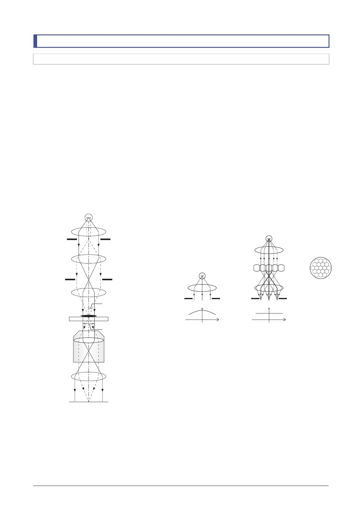
BF microscopy optical system
The optical system of BF microscopy with Köhler
illumination is shown in the following figure.
Köhler illumination optical system
Light from the light source is converted by a collector
lens into a parallel luminous flux and passes the field
diaphragm. Light emitted from the aperture
diaphragm is concentrated by a field lens and an
image of the light source is formed at the aperture
diaphragm.
Light emitted from the aperture diaphragm is
converted by the condenser lens into a parallel
luminous flux and illuminates the specimen. The field
diaphragm can restrict the illumination range of the
specimen plane. Unnecessary light can be cut and
thus a clear field of view free from flare or ghost can
be obtained. The image of the light source can be
restricted by the aperture diaphragm. This changes
the brightness of the field of view. The incidence
angle θ of the illumination also changes. Because of
this incidence angle θ, adjustment of the aperture
diaphragm causes the contrast resolution and focal
depth of images to change.
Use of fly-eye lens to make illumination uniform
The uniformity of the illumination can be further
increased using fly-eye lenses for the illumination
attachment. A fly-eye lens is an array of small
hexagonal lenses. The name “fly-eye lens” derives
from the fact that its shape is similar to the
compound eye of a fly. The following figure explains
the effects of a fly-eye lens.
Use of fly-eye lens to make illumination uniform
Generally speaking, light emitted obliquely from the
light source has a lower brightness than light emitted
vertically from the light source (light distribution
characteristic). If no fly-eye lens is used, light emitted
vertically from the light source irradiates the center of
the field of view, and light emitted obliquely from the
light source irradiates the periphery of the field of
view. As a result, light intensity in the periphery of the
field of view is lower than in the center of the field
even in the case of Köhler illumination. On the other
hand, if a fly-eye lens is used, light beams of
different emergence angles are intermixed and
irradiate their respective position in the field of view.
As a result, the field of view can be illuminated with a
uniform intensity regardless of the light distribution
characteristics of the light source.
Light source
Collector lens
Field diaphragm
Field lens
Aperture diaphragm
Condenser lens
Specimen
Objective
2nd tube lens
Image plane
ngle of incidence θ
Illumination range
Light source
Collector
lens
Fly-eye
lens
Field
diaphragm
Light source
Collector
lens
Field
diaphragm
Illuminance distribution
on the field diaphragm plane
Illuminance distribution
on the field diaphragm plane
Dark Bright Dark Bright Bright Bright