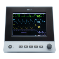
Patient Monitor User Manual Monitoring ECG
- 74 -
5 consecutive ventricular beats, and 40 bpm ≤ ventricular HR < 100 bpm.
No QRS is detected within the heartbeat pause threshold value that has
been set.
The measurement value of Pause/min is greater than high alarm limit that
has been set.
The delayed ventricular beats detected in normal heartbeats occur more
than or equal to 3 times within 30 s.
Different forms of ventricular premature beats are detected in 15 beats.
The single ventricular premature beat between 2 sinus beats with normal
interval occurs more than or equal to 3 times within 30 s.
The dominant rhythm of N, A, N, A, N, A, and the rhythm number
exceeds the number of threshold value that has been set (N =
supraventricular beat, A = atrial beat).
The dominant rhythm of N, N, A, N, N, A, N, N, A, and the rhythm
number exceeds the number of threshold value that has been set.
The signal amplitudes of I, II and III leads shall not exceed alarm
threshold value that has been set. PS: this alarm is available for 5, 6 or 10
electrodes only, not available for 3 electrodes.
Selecting an ECG lead for Arrhythmia:
In arrhythmia monitoring, it is important to select the appropriate lead.
For non-paced patients, the guidelines are:
- QRS should be tall and narrow (recommended amplitude > 0.5 mV)
- R wave should be above or below the baseline (but not biphasic)
- T wave should be smaller than 1/3 of the R wave height
- P wave should be smaller than 1/5 of the R wave height.
For paced patients, in addition to above guidelines, the pacemaker signal should also:
- Not wider than normal QRS
- The QRS complexes should be at least twice the height of the pacing pulse
- Large enough to be detected, without repolarization signal
According to Standard ISO60601-2-27, the minimum detection level of the QRS complex is set
to 0.15 mV, to prevent the detection of P-wave or baseline noise as QRS complexes. Adjusting
ECG displayed waveform size (gain adjustment) won’t influence ECG signals which are used for
arrhythmia analysis. If the ECG signal is too small, a false asystole alarm may occur.
Aberrantly-Conducted Beats:
As not recognizing the P waves, the monitoring system is difficult to distinguish between
aberrantly-conducted beats and ventricular heartbeat. If the aberrantly-conducted beat is similar
to ventricular tachycardia, it may be classified as ventricular. Make sure to select such a lead, the
aberrantly-conducted beats have an R wave that is as narrow as possible to minimize the incorrect