Image Optimization 5-11
HScale
Description Display or hide the width scale (horizontal scale).
The scale of the horizontal scale is the same as that of vertical scale
(depth), they change together in zoom mode, or when the number of the
image window changes. When image is turned up/down, the HScale will
also be inverted.
Operation Click [HScale] on the soft menu to display or hide the scale.
5.4 M Mode Image Optimization
5.4.1 M Mode Exam Protocol
1. Select a high-quality image during B mode scanning, and adjust to place the area of
interest in the center of the B mode image.
2. Press <M> on the control panel, and roll the trackball to adjust the sampling line.
3. Press <M> on the control panel again or <Update> to enter M mode, and then you
can observe the tissue motion along with anatomical images of B mode.
During the scanning process, you can also adjust the sampling line accordingly when
necessary.
4. Adjust the image parameters to obtain optimized images.
5. Perform other operations (e.g. measurement and calculation) if necessary.
If you choose "Enter 1D Mode Directly" in "[Setup]→[Image Preset]→[Other]", then
the sampling line will be displayed at all times in B mode images, and pressing <M>
will directly enter M mode.
5.4.2 M Mode Parameters
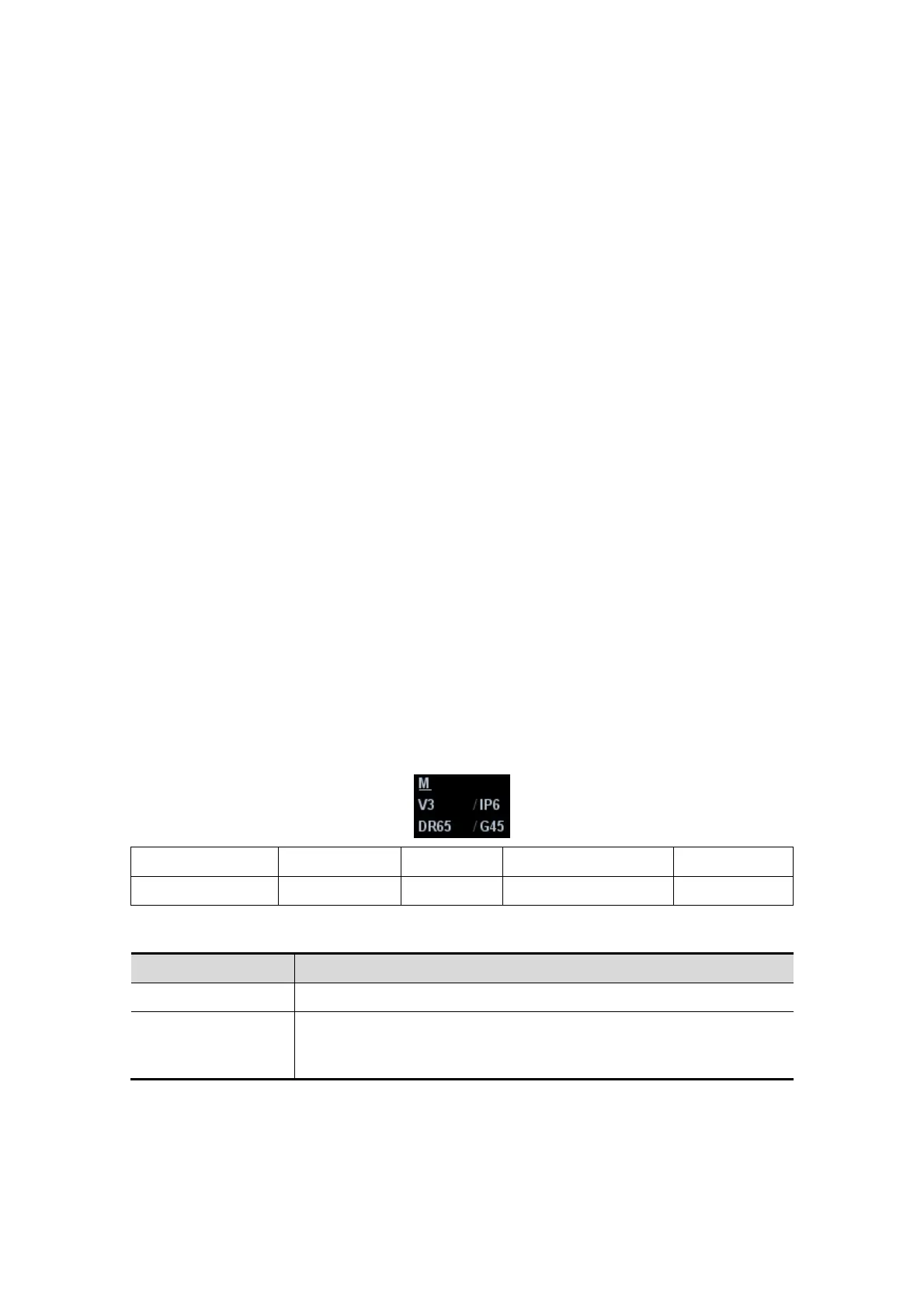
In M mode scanning, the image parameter area in the upper left corner of the screen
displays the real-time parameter values as follows:
Display V 3 IP 6 DR 65 G 45
Parameter M Speed M IP M Dynamic Range M Gain
Parameters that can be adjusted to optimize the M mode image are indicated in the
following.
Adjustment Items
Control Panel Gain, TGC, Depth
Menu and Soft
Menu
IP, Time Mark, Speed, Colorize, Colorize Map, Acoustic Power,
Edge Enhance, Frequency, Gray Map, Focus Position, Dynamic
Range, M Soften, Curve, Gray Rejection, γ, Display Format