Table of contents
4 / 106 Instruction Manual Keratograph 5M (G/77000/XXXX/DE – Rev05)
10.8 Manual measurement.........................................................................36
10.9 Image Capture with the Foot Switch ............................................36
10.10 Finishing the Exam...............................................................................36
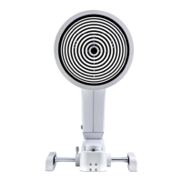
11 Performing a “Topography” Examination ................................................37
11.1 Patients who are sensitive to light: Low glare with white flash
38
11.2 Manually marking the center of the Placido rings ..................38
12 Contact lens back side measurement........................................................40
12.1 Parts for Measuring the Back Surfaces of Contact Lenses ...40
12.2 Fill the Contact Lens Holder with Water......................................40
12.3 Measure the Back Surface of the Dry Contact Lens................41
12.4 Fixating the Contact Lens..................................................................41
12.5 Fasten the Mounted Contact Lens Holder into Place ............41
12.6 Fully assembled contact lens holder.............................................41
12.7 CL Back Surface.....................................................................................42
13 Performing a “TF Scan” Examination .........................................................43
13.1 Examination of the Lipid Layer........................................................44
13.2 TF Dynamics Examination .................................................................45
13.3 Measuring the Tear Meniscus Height...........................................46
13.4 Measuring NIKBUT...............................................................................47
14 Performing an “R Scan” Examination.........................................................49
15 Performing a “Meibo Scan” Examination .................................................50
15.1 Upper and Lower Eyelid Image.......................................................50
15.2 Recording a Single Image.................................................................51
16 Performing a “Pupillometry” Examination ...............................................52
16.1 Adjustment..............................................................................................53
16.2 Measuring values display ..................................................................53
16.3 Pupillogram ............................................................................................54
16.4 Asymmetric Test....................................................................................54
16.5 Manually...................................................................................................54
17 Imaging .................................................................................................................55
17.1 Recording a Fluo Image.....................................................................56
17.2 Near Portion Height Measurement...............................................57
17.3 Eyelid Angle Measurement...............................................................58
17.4 New Recording......................................................................................59
17.5 Adjusting Illumination, Magnification Changer
and Camera.............................................................................................59
17.5.1 Adjust Illumination: [Illumination] groupbox ..........60
17.5.2 Magnification changer.....................................................60
17.5.3 Adjusting the Camera: Camera groupbox................60
17.5.4 Buttons...................................................................................61
17.5.5 Selecting and saving settings........................................61
17.5.6 Using your own settings for an image program....61
18 Performing Dry Eye Examinations: JENVIS Dry Eye Report...............62
18.1 Selecting the Exam Type....................................................................63
18.2 Performing the selected examination ..........................................63
18.3 Filling in the “Recommendation” field .........................................64
18.3.1 Using Text Blocks ...............................................................64
18.3.2 Entering own Texts ............................................................64
18.3.3 Deleting Texts......................................................................64
18.4 Printing the JENVIS Dry Eye Report ..............................................64
18.5 DEQ OSDI ................................................................................................65