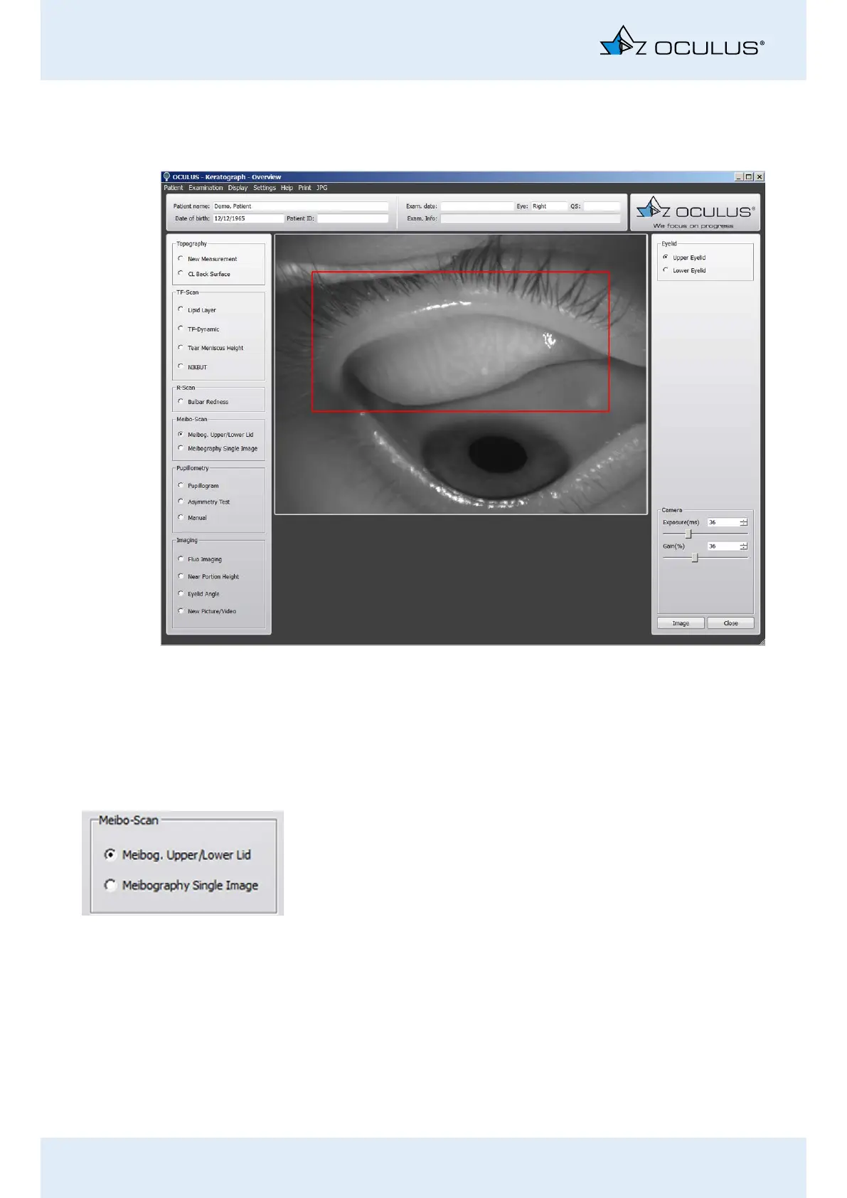
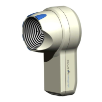
15 Performing a “Meibo Scan” Examination
50 / 106 Instruction Manual Keratograph 5M (G/77000/XXXX/EN – Rev05)
15 Performing a “Meibo Scan” Examination
This examination visualizes the meibomian glands. They are displayed
three-dimensionally. You can make images of the upper and lower eye-
lid as well as individual images. Changes can be viewed and classified.
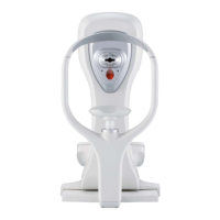
15.1 Upper and Lower Eyelid Image
Enable the [Meibog. Upper/Lower Lid] radio button in the right
group box [Meibo-Scan].
Ectropionize the upper eyelid first.
Adjust the camera if necessary, Chap. 17.5, page 59.
Position the camera so that the upper eyelid fits in the red-framed
recording box.
Focus on the Meibom glands.
Start the recording of the upper eyelid. To do this, press the [Im-
age] button.
Use the foot switch alternatively, (Chap. 10.10, page 36).
Repeat the steps for the lower eyelid.
Fig. 15-1: Meibo-Scan examinations